Q.Draw DNA model of Watson-Crick. (with only two labelling) OR Write the tertiary structure of protein.
You're viewing a preview — the full solution, concept, methods & PYQ mapping are locked.
Start your 14-day free trial to unlock the full solution →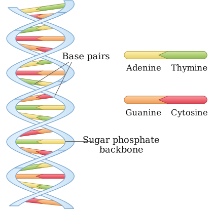
The Watson-Crick DNA model describes DNA as a right-handed double helix: two sugar-phosphate backbones running antiparallel, connected internally by hydrogen-bonded, complementary nitrogenous base pairs (A pairs with T by two hydrogen bonds; G pairs with C by three hydrogen bonds).
A hand-drawn diagram of the Watson-Crick DNA model would show:
-
Two long, twisted ribbon-like strands running side by side in a right-handed helix, representing the sugar-phosphate backbones (each made of alternating deoxyribose sugar and phosphate groups linked by phosphodiester bonds) — this is Label 1: 'Sugar-phosphate backbone'.
-
The two strands run in opposite (antiparallel) directions — one strand 5' to 3' from top to bottom, the other 3' to 5' from top to bottom.
-
Connecting the two backbones like the rungs of a twisted ladder are pairs of nitrogenous bases projecting inward, each pair joined by hydrogen bonds: adenine always pairs with thymine (2 hydrogen bonds) and guanine always pairs with cytosine (3 hydrogen bonds) — this is Label 2: 'Nitrogenous base pairs (A-T, G-C) joined by hydrogen bonds'.
…
Unlock everything free for 14 days
- Full step-by-step solutions
- Concept-first explanations
- Methods, shortcuts & mistakes
- PYQ mapping + timed mock tests
Full access for 14 days. No credit card required.