Q.Describe, with the help of a diagram, the development of a mature embryo sac from a megaspore mother cell in angiospermic plants. [FIGURE: mature embryo sac development — not reproducible as text]
[FIGURE: three diagrams — not reproducible as text]
You're viewing a preview — the full solution, concept, methods & PYQ mapping are locked.
Start your 14-day free trial to unlock the full solution →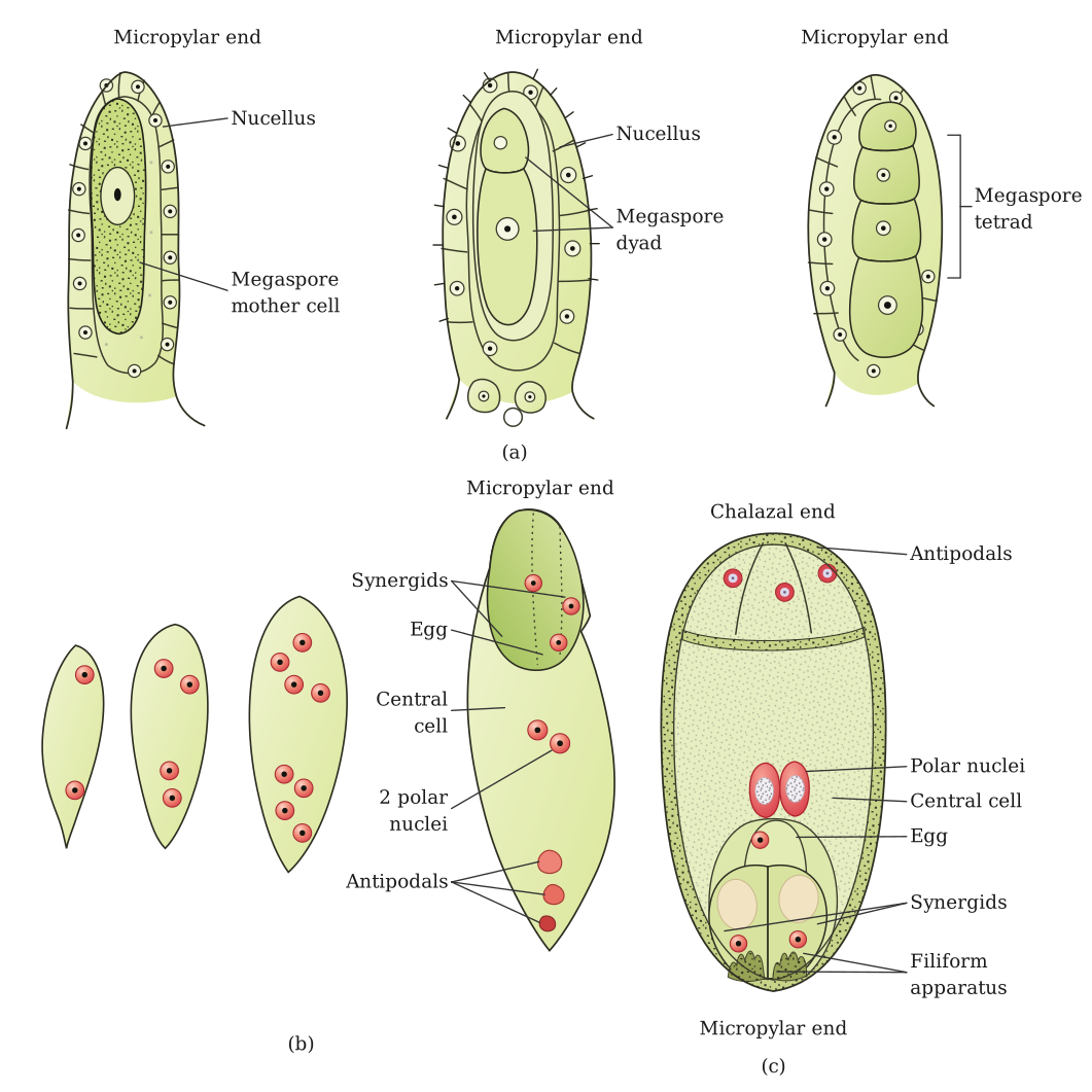
The 8-nucleate, 7-celled embryo sac (Polygonum-type) develops from the single surviving megaspore by three rounds of mitosis.
[FIGURE: Development of the mature (monosporic, Polygonum-type) embryo sac.] The megaspore mother cell (MMC), located within the nucellus of the ovule, undergoes meiosis (meiosis I followed by meiosis II) to produce a linear tetrad of four haploid megaspores. Of these four, usually only the megaspore situated at the chalazal end remains functional, while the other three degenerate.
The functional megaspore then enlarges and undergoes three successive free-nuclear mitotic divisions: the first division gives a 2-nucleate embryo sac, the second gives a 4-nucleate stage, and the third gives an 8-nucleate embryo sac. Following this, cell walls are laid down and the eight nuclei get organized: three nuclei move to the micropylar end and form the egg apparatus — one egg cell flanked by two synergids; three nuclei move to the chalazal end to form three antipodal cells; and the remaining two nuclei (one from each pole), called polar nuclei, move to the centre and lie in a common cytoplasm (the central cell), often fusing to form the diploid secondary/definitive nucleus. Thus the typical mature embryo sac, though 8-nucleate, is organizationally 7-celled.
OR:
[FIGURE — three separate labelled diagrams, not reproducible as text:]
(a) Mature microspore — a two-celled pollen grain at shedding stage, showing a large vegetative (tube) cell and a smaller generative cell lying within it, enclosed by the two-layered pollen wall (exine with germ pores, and intine). …
Unlock everything free for 14 days
- Full step-by-step solutions
- Concept-first explanations
- Methods, shortcuts & mistakes
- PYQ mapping + timed mock tests
Full access for 14 days. No credit card required.