Q.What is stomatal apparatus? Explain the structure of stomata with a labelled diagram.
🔒You're viewing a preview — the full solution, concept, methods & PYQ mapping are locked.
🔒 Start your 14-day free trial to unlock the full solution →Concept understanding — Monocot Dicot Anatomy
Monocot vs Dicot Anatomy – A First Look
Imagine you are holding two seeds: a grain of rice and a kidney bean. Soak them overnight. The rice grain splits open easily along one line — it has a single seed leaf. The bean splits into two halves — it has two seed leaves. That single versus double seed leaf is the root of the entire distinction: monocot (one cotyledon) versus dicot (two cotyledons).
But the difference doesn't stop at the seed. It runs through the entire plant — root, stem, leaf, and flower — like a blueprint that shapes how the plant grows, transports water, and arranges its parts.
The Core Idea
Monocots and dicots are two major groups of flowering plants (angiosperms) that differ in the arrangement of vascular tissue, number of cotyledons, leaf venation, root system, and floral parts.
The word "cotyledon" means seed leaf — the first leaf that emerges from a seed. Monocots have one; dicots have two. Everything else follows from that developmental difference.
The Five Key Differences at a Glance
| Feature | Monocot | Dicot |
|---|---|---|
| Cotyledons | One | Two |
| Leaf venation | Parallel (veins run side by side) | Reticulate (veins form a net) |
| Root system | Fibrous (many thin roots) | Taproot (one main root) |
| Vascular bundles in stem | Scattered | Arranged in a ring |
| Floral parts | Multiples of 3 | Multiples of 4 or 5 |
Let's unpack each one.
1. Leaf Venation – The First Clue
Pick up a leaf of grass (monocot) and a leaf of a mango tree (dicot). Run your finger along the veins.
- Monocot leaf: Veins run parallel to each other from base to tip, like train tracks. Example: bamboo, wheat, corn.
- Dicot leaf: Veins branch out from a central midrib, forming a net-like pattern. Example: rose, sunflower, neem.
Why? The vascular bundles (xylem and phloem) in monocot leaves are arranged in parallel rows, while in dicots they branch repeatedly. This is the easiest way to tell them apart without a microscope.
2. Root System – Underground Architecture
Dig up a small grass plant and a small bean plant. Rinse the soil.
- Monocot root: A tangled mass of thin, branching roots of roughly equal size — fibrous root system. No single root dominates. This anchors the plant well in topsoil and prevents erosion.
- Dicot root: One thick main root (the taproot) grows straight down, with smaller lateral roots branching off. This reaches deep water and stores food (think carrot, radish).
If you see a plant with a taproot, it is almost certainly a dicot. Fibrous roots are a strong indicator of a monocot — but there are exceptions (e.g., some dicots also have fibrous roots in waterlogged soil).
3. Stem – The Internal Arrangement
This is where the anatomy gets precise. Cut a thin cross-section of a stem and stain it. Under a microscope, you see the vascular bundles — clusters of xylem (water) and phloem (food).
- Monocot stem: Vascular bundles are scattered throughout the ground tissue, with no particular order. There is no distinct cortex or pith. Example: maize, sugarcane.
- Dicot stem: Vascular bundles are arranged in a ring around a central pith. This ring separates the outer cortex from the inner pith. This ring arrangement allows for secondary growth (increase in girth) — which is why dicot trees can grow thick trunks, while monocot palms cannot.
Monocot stem: Scattered vascular bundles, no secondary growth.
Dicot stem: Ring of vascular bundles, secondary growth possible.
4. Floral Parts – Counting Petals
Look at a flower. Count the petals, sepals, stamens.
- Monocot flower: Parts in multiples of 3. Example: lily (3 petals, 3 sepals, 6 stamens), orchid, grass flowers.
- Dicot flower: Parts in multiples of 4 or 5. Example: rose (5 petals), mustard (4 petals), sunflower (5 petals).
This is not a hard rule — some dicots have 3-merous flowers — but it holds for the vast majority.
5. The Seed – Where It All Begins
Soak a corn grain (monocot) and a bean seed (dicot). Split them open.
- Monocot seed: One cotyledon. The food is stored in a separate tissue called the endosperm. The cotyledon absorbs and transfers food to the embryo. …
The stomatal apparatus is the whole functional unit of a stoma — the stomatal pore together with the two guard cells and, in some plants, the neighbouring subsidiary cells. Stomata regulate transpiration and gaseous exchange.
Structure of a stoma:
- Each stoma is a minute pore in the epidermis, bounded by two guard cells.
- In dicots the guard cells are bean-shaped (kidney-shaped); in grasses (monocots) they are dumb-bell shaped.
- The inner walls of the guard cells (towards the pore) are thick and inelastic, while the outer walls are thin and elastic.
- Guard cells contain chloroplasts and control opening/closing by changes in their turgidity.
- Subsidiary (accessory) cells may surround the guard cells. …
The stomatal apparatus is the stomatal pore plus its guard cells and subsidiary cells, and each stoma opens and closes through turgor-driven shape changes of unequally-thickened guard cells.
What the stomatal apparatus is
The stomatal apparatus is the complete structural and functional unit associated with a stoma. It comprises:
- the stomatal aperture (pore),
- the two guard cells that bound the pore, and
- the surrounding subsidiary (accessory) cells present in many plants.
Stomata occur mainly in the epidermis of leaves (and young stems) and are responsible for transpiration and the exchange of gases (carbon dioxide and oxygen) between the plant and the atmosphere.
Structure of a stoma
- Each stoma is a small pore surrounded by two specialised epidermal cells called guard cells.
- The guard cells are bean-shaped (kidney-shaped) in most dicots, and dumb-bell shaped in grasses and other monocots.
- Unlike other epidermal cells, guard cells possess chloroplasts.
- The wall of a guard cell is unevenly thickened: the wall facing the pore (inner wall) is thick and inelastic, while the outer wall is thin and elastic.
- When the guard cells absorb water and become turgid, the thin outer walls stretch more, curving the cells outward and opening the pore; when they lose water and become flaccid, the pore closes.
- Subsidiary cells may be arranged around the guard cells and help in the opening and closing mechanism. …
Method: Reasoning from Mechanics, Not Memorising the Parts List
Rather than listing the stomatal apparatus's components and moving on, you can arrive at (and remember) the same structure by asking a single mechanical question: what shape would a pressure-driven valve need, to open a gap without any moving hinge or muscle?
Start from the constraint: a stoma has no muscle and no external lever — it must open and close using only water pressure (turgor) inside its own two guard cells.
- If a guard cell's wall were evenly thick all around, swelling with water would just make it a bigger, rounder cell — it would push outward uniformly and would not reliably pull away from its neighbour to create a gap.
- To force the cell to bow away from the pore specifically, the wall touching the pore must resist stretching more than the wall facing away from it. That is exactly why the inner (pore-side) wall is thick and the outer wall is thin: when turgor pressure rises, the thin outer wall stretches more than the thick inner wall can, and this uneven stretching bends the whole cell into a curve — bowing the two guard cells apart and opening the pore between them.
- When turgor falls, the cells relax back toward their unstressed (straighter) shape, and the pore closes. …
- AP EAPCET 2025Set ap-2025-05-20-FN1 markMCQQ.Match the following : List - I / List - II A. Polyarch xylem - I. Bulliform cells B. Colourless cells in epidermis - II. Cork cells C. Ring arrangement of vascular bundles - III. Monocot root D. Suberin deposition on cell wall - IV. Dicot stem (A) A-IV, B-III, C-I, D-II (B) A-III, B-II, C-I, D-IV (C) A-III, B-I, C-IV, D-II (D) A-III, B-IV, C-I, D-II
›Reveal solutionSolution
Matching anatomical features to their tissue/organ context gives A-III (monocot root), B-I (bulliform cells), C-IV (dicot stem), D-II (cork cells) — option (C).
Concept and Intuition
Each listed feature is a diagnostic anatomical marker used to identify a specific tissue or organ: xylem arch number for root type, colourless epidermal cells for grass-leaf water-loss control, vascular bundle arrangement for stem type, and suberin deposition for the cork layer.
Step-by-Step Solution
- A) Polyarch xylem (many xylem groups arranged around the periphery) is a defining feature of the monocot root — III.
- B) Colourless cells in the epidermis refers to the large, vacuolated, colourless bulliform cells found in the upper epidermis of grass leaves, involved in leaf rolling/unrolling to reduce water loss — I.
- C) Ring arrangement of vascular bundles (a single ring of collateral, open bundles) is characteristic of the dicot stem — IV. …
- AP EAPCET 2024Set ap-2024-05-16-FN1 markMCQQ.Identify the Anatomical structures based on these following characters given in a series • Lysigenous cavities in the vascular bundles • Conjoint, open and endarch protoxylem in vascular bundles • More number of stomata in abaxial epidermis (A) Monocot stem, Dicot stem, Dicot Leaf (B) Monocot root, Dicot root, Dicot Leaf (C) Monocot Leaf, Dicot Leaf, Dicot stem (D) Dicot stem, Dicot root, Monocot Leaf
›Reveal solutionSolution
The three clues identify, in order, a monocot stem (lysigenous protoxylem lacuna), a dicot stem (open, conjoint, endarch vascular bundles), and a dicot leaf (more abaxial stomata). Answer: (A).
Concept and Intuition
Plant anatomy questions like this test whether you can recognise a tissue from its functional signature rather than just memorising a list.
- A lysigenous cavity forms when cells break down (lyse) to create a cavity. In monocot stems (grasses like maize), the protoxylem elements are the first, narrowest vessels laid down; as the internode elongates, these delicate elements get torn apart and their debris dissolves, leaving a fluid-filled cavity called the protoxylem lacuna — this is a textbook diagnostic feature of monocot stem vascular bundles.
- "Open" vascular bundles contain a strip of cambium between xylem and phloem, allowing secondary growth; this is present only in dicot stems (and absent in monocot stems, whose bundles are "closed"). Both monocot and dicot stems have endarch xylem (protoxylem towards the centre, metaxylem towards the periphery), but the presence of cambium (open condition) is what pins this down specifically to a dicot stem.
- Stomatal distribution reflects orientation to light and gravity: dorsiventral (dicot) leaves lie horizontally, so the upper (adaxial) surface faces more sun and has fewer stomata (to reduce water loss), while the lower (abaxial) surface, shaded and cooler, carries more stomata. Isobilateral (monocot) leaves, often oriented vertically, show roughly equal numbers on both surfaces.
Step-by-Step Solution
- "Lysigenous cavities in the vascular bundles" ⇒ Monocot stem. …
- AP EAPCET 2023Set ap-2023-05-22-AN1 markMCQQ.Match the following List I: I) Well developed large pith, II) Casparian strips, III) Scelerenchymatous bundle sheath, IV) Endarch protoxylem List II: A) Endodermis, B) Monocot stem, C) Dicot stem, D) Monocot root (A) I-B, II-A, III-D, IV-C (B) I-D, II-A, III-B, IV-C (C) I-D, II-C, III-A, IV-B (D) I-C, II-A, III-D, IV-B
›Reveal solutionSolution
Matching anatomical features to their tissue location: large pith → monocot root; Casparian strips → endodermis; sclerenchymatous bundle sheath → monocot stem; endarch protoxylem → dicot stem — giving I-D, II-A, III-B, IV-C.
Concept and Intuition
This tests recall of distinguishing internal anatomical features across the four classic plant-anatomy categories (monocot/dicot × root/stem), each with hallmark structural signatures used to identify a cross-section under the microscope.
Step-by-Step Solution
- I. Well developed large pith: dicot roots typically have small/absent pith (xylem often extends toward the centre), whereas monocot roots are polyarch with a prominent, large parenchymatous pith — so I matches D) Monocot root.
- II. Casparian strips: these are suberized bands present specifically in the endodermis (the innermost cortical layer, in both monocot and dicot roots) — so II matches A) Endodermis.
- III. Sclerenchymatous bundle sheath: each scattered vascular bundle in a monocot stem is characteristically surrounded by a sclerenchymatous bundle sheath cap — so III matches B) Monocot stem. …
- AP EAPCET 2023Set ap-2023-05-22-AN1 markMCQQ.The hypodermis tissues in dicot and monocot stems respectively (A) Collenchyma and Sclerenchyma. (B) Sclerenchyma and Collenchyma. (C) Sclerenchyma in both. (D) Collenchyma in both.
›Reveal solutionSolution
Dicot stems have a collenchymatous hypodermis (living, flexible mechanical support); monocot stems have a sclerenchymatous hypodermis (dead, rigid support) — matching option (A).
Concept and Intuition
The hypodermis is the layer of ground tissue immediately below the epidermis, and its composition differs consistently between the two major angiosperm groups, reflecting their different overall stem mechanics (dicot stems can grow thicker via secondary growth and rely on flexible living support early on; monocot stems, lacking secondary growth, rely on permanent rigid support from the start).
Step-by-Step Solution
- Dicot stem (e.g. sunflower, Helianthus) cross-section: epidermis → hypodermis of collenchyma (living cells with extra cellulose thickening at the corners, giving mechanical strength while allowing some flexibility and growth) → cortex. …
- AP EAPCET 2023Set ap-2023-05-22-FN1 markMCQQ.Select the incorrect statements related to anatomical characters from the following I) Endodermis is a part of the stele. II) Endodermis with casparian strips are found in the dicot stem. III) Bulliform cells are found in the upper epidermis of the isobilateral leaf. IV) Vascular bundles are open in monocot stems. (A) I, II, IV (B) I, II, III (C) II, IV (D) III, IV
›Reveal solutionSolution
Endodermis is part of the cortex (not stele) and its prominent casparian strips are a root feature (not typical of stems); monocot stem bundles are closed, not open — so I, II and IV are the incorrect statements, while III (bulliform cells in isobilateral leaves) is correct.
Concept and Intuition
Plant anatomy assigns each tissue layer to a specific zone: cortex (hypodermis, general cortex, endodermis) lies outside the stele (pericycle + vascular tissue + pith). The endodermis, though it borders the stele, is conventionally classified as the innermost cortical layer, not part of the stele itself. Casparian strips — waxy suberin bands that force water/solutes through the symplast — are the textbook hallmark of ROOT endodermis; dicot stems' endodermal layer (often called the starch sheath, due to abundant starch grains) is not typically distinguished by prominent casparian strips. Isobilateral (monocot, e.g., grass) leaves have bulliform/motor cells in their upper epidermis that help the leaf roll and unroll to reduce water loss. Vascular bundles in monocot stems are "closed" (no cambium, cannot add secondary growth), whereas dicot stems have "open" bundles (with cambium).
Step-by-Step Solution
- I) Endodermis is the innermost cortical layer, not a stele component → statement is INCORRECT.
- II) Casparian strips are the signature feature of root endodermis; dicot stem endodermis (starch sheath) is not typically defined by casparian strips → statement is INCORRECT. …
- AP EAPCET 2023Set ap-2023-05-23-FN1 markMCQQ.Identify the common character present in both dicot and monocot stems (A) Pericycle and Endodermis. (B) Open vascular bundles. (C) Well developed large pith. (D) Conjoint vascular bundles.
›Reveal solutionSolution
Identify the one anatomical feature genuinely shared between dicot and monocot stems, despite their many differences.
Concept and Intuition
Dicot and monocot stems differ in vascular bundle arrangement (ring vs scattered), in whether bundles are open (with cambium, dicot) or closed (without cambium, monocot), and in pith development (well-developed in dicot, often not distinct in monocot). What they DO share is that in both, the vascular bundles are conjoint — xylem and phloem are located together within the same bundle (collateral arrangement), rather than as separate, distinct bundles.
Step-by-Step Solution
- Pericycle and endodermis: prominent/distinct in dicot stem, but not clearly differentiated in monocot stem — not a common feature.
- Open vascular bundles: true only for dicot stems (which have cambium); monocot bundles are closed — not common. …
- AP EAPCET 2021Set ap-2021-09-03-AN1 markMCQQ.The characters restricted to monocot Stem & Leaf respectively ________ (A) Trichomes & Bulliform cells (B) Lysigenous cavity & Bulliform cells (C) Trichomes & Lysigenous cavity (D) Bulliform cells & Lysigenous cavity
›Reveal solutionSolution
This tests which anatomical features are unique to monocot stems versus monocot leaves. The answer is (B).
Concept and Intuition
Monocot (grass-type) anatomy has a few textbook 'signature' features that examiners like to separate by organ. In the stem, the vascular bundles are scattered, each with a sclerenchymatous bundle sheath, and — distinctively — the protoxylem elements often rupture during growth, leaving behind a large, water-filled lysigenous cavity in the centre of the bundle/stem. This is a stem-specific feature not seen in monocot leaves.
In the leaf, the isobilateral monocot leaf has large, vacuolated, thin-walled bulliform cells on the upper (adaxial) epidermis. These cells lose turgor under water stress, causing the leaf to roll inward and reduce transpiring surface — a leaf-specific adaptation.
Trichomes (hair-like epidermal outgrowths) are found generally on both monocot and dicot stems/leaves, so they cannot be the 'restricted-to-monocot' answer for either organ.
Step-by-Step Solution …
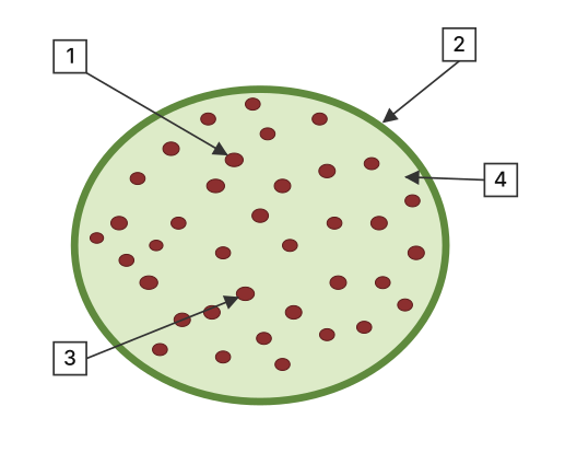
- AP EAPCET 2021Set ap-2021-09-06-FN1 markMCQQ.Identify the labeled parts 1, 2, 3, 4 in the below figure? [FIGURE] (a circular cross-section of a plant organ with four arrows pointing to labelled boxes 1, 2, 3, 4 marking different tissue layers from the outer edge inward) (A) 1 - Ground tissue, 2 - Vascular bundles, 3 - Epidermis, 4 - Hypodermis (B) 1 - Epidermis, 2 - Hypodermis, 3 - Vascular bundles, 4 - Ground tissue (C) 1 - Vascular bundles, 2 - Epidermis, 3 - Ground tissue, 4 - Hypodermis (D) 1 - Hypodermis, 2 - Epidermis, 3 - Vascular bundles, 4 - Ground tissue
›Reveal solutionSolution
The scattered (not ring-arranged) bundles mark this as a monocot stem cross-section: outer rim = Epidermis, just inside it = Hypodermis, the discrete bodies = Vascular bundles, and the surrounding bulk tissue = Ground tissue.
Concept and Intuition
In a monocot stem, unlike a dicot stem, there is no distinct cortex/pith division — vascular bundles are scattered throughout an undifferentiated ground tissue (a mix of parenchyma). Working from the outside in: the single outer layer of cells is the epidermis; immediately beneath it is a narrow band of sclerenchymatous hypodermis providing mechanical strength; the rest of the interior, filled with parenchymatous ground tissue, has numerous discrete vascular bundles scattered through it (denser near the periphery).
Step-by-Step Solution
- Identify the outermost boundary of the cross-section — label '2' — as the Epidermis.
- The layer just inside the epidermis — label '4' — as the Hypodermis (mechanical layer adjoining the epidermis).
- A discrete, distinctly-outlined body embedded well inside the section — label '1' — as one of the scattered Vascular bundles. …
- AP EAPCET 2021Set ap-2021-09-06-FN1 markMCQQ.Select the mismatch pair from the following? (A) Monocot root – polyarchy (B) Monocot stem – Lysigenous cavity (C) Dicot leaf – Bulliform cells (D) Dicot stem – Sclerenchymatous pericycle
›Reveal solutionSolution
Bulliform cells are a diagnostic feature of monocot (grass-type) leaves, not dicot leaves, making (C) the mismatched pair.
Concept and Intuition
Anatomical identification questions hinge on knowing which features are exclusive to monocots vs dicots. Polyarchy (many xylem strands) is typical of monocot roots because they have a wider stele with numerous vascular strands. Lysigenous cavities forming by breakdown of central pith cells are seen in many monocot stems (e.g., grasses, maize). Sclerenchymatous pericycle (a mechanical cap over vascular bundles/protecting the stele) is typical of dicot stems. Bulliform cells, however, are large, thin-walled, empty epidermal cells found on the upper epidermis of grass (monocot) leaves — they lose water and shrink to fold the leaf during water stress, a mechanism absent from dicot leaf epidermis.
Step-by-Step Solution
- Verify (A): monocot root – polyarchy → TRUE, matches textbook anatomy.
- Verify (B): monocot stem – lysigenous cavity → TRUE, seen in maize/grass stems. …
🎓Unlock everything free for 14 days
- ✓Full step-by-step solutions
- ✓Concept-first explanations
- ✓Methods, shortcuts & mistakes
- ✓PYQ mapping + timed mock tests
Full access for 14 days. No credit card required.