Q.Make a chart (with diagrammatic representation) showing a restriction enzyme, the substrate DNA on which it acts, the site at which it cuts DNA and the product it produces.
Concept understanding — Restriction Enzyme Action
Imagine you have a long, tangled piece of string, and you need to cut it into smaller, specific pieces — not just anywhere, but exactly at the places where a certain pattern of letters appears. That is the core idea behind restriction enzyme action.
In the world of biology, the "string" is a DNA molecule — the long, thread-like chemical that carries the genetic instructions for every living thing. A restriction enzyme is a molecular "scissors" that cuts DNA, but it is incredibly precise. It does not chop randomly. Instead, it recognises a very specific, short sequence of DNA letters (usually 4 to 8 base pairs long) and cuts only at that exact spot.
Think of it like a word processor's "Find and Replace" function, but instead of replacing text, the enzyme finds a specific word and cuts the page at that word.
This ability to cut DNA at precise locations is what makes restriction enzymes the fundamental tool of genetic engineering. Without them, scientists would have no way to isolate a specific gene from a long DNA strand.
How does the enzyme "know" where to cut?
The DNA molecule is made of two strands twisted together (the famous double helix). Each strand has a sequence of four chemical "letters": A, T, G, and C. A restriction enzyme scans along the DNA until it finds its target sequence — a short, palindromic pattern (meaning it reads the same forwards on one strand and backwards on the other). For example, the enzyme EcoRI recognises the sequence GAATTC.
When it finds this exact sequence, it binds to the DNA and makes a cut in both strands. The cut can be one of two types:
- Sticky ends: The enzyme cuts the two DNA strands at different points, leaving short, single-stranded overhangs. These overhangs are like pieces of Velcro — they can easily stick to a complementary overhang from another DNA piece cut by the same enzyme. This is extremely useful for joining different DNA fragments together.
- Blunt ends: The enzyme cuts both strands straight across at the same point, leaving no overhang. These are harder to join together later, but they are still useful.
Why does this matter?
Restriction enzymes are the reason we can manipulate DNA at all. They allow scientists to:
- Cut out a specific gene from the DNA of one organism (say, the human insulin gene).
- Cut open a carrier DNA (like a plasmid from a bacterium) at the same spot.
- Insert the gene into the carrier, because the sticky ends match perfectly.
- Splice the carrier back together using another enzyme (DNA ligase), creating a recombinant DNA molecule.
This is the foundation of modern biotechnology — from producing human insulin in bacteria to creating genetically modified crops and developing gene therapies.
The key takeaway from the NCERT textbook is that restriction enzymes are molecular scissors that cut DNA at specific recognition sites. Their action produces fragments with either sticky ends or blunt ends, and this precise cutting is what makes genetic engineering possible. The enzyme itself is a protein, and it is named after the bacterium from which it is isolated (e.g., EcoRI from Escherichia coli).
In short: Restriction enzyme action is the controlled, precise cutting of DNA at predetermined locations — the first and most essential step in any DNA manipulation experiment.
Restriction enzyme action is one of the most tested mechanisms in the NCERT Class 12 Biology chapter on Biotechnology: Principles and Processes, and appears in searches like "restriction enzymes class 12 biology sticky ends" or "EcoRI recognition site important questions." This is a near-certain topic in both CBSE board papers and NEET's biotechnology section every year.
Restriction enzymes are molecular scissors that cut DNA at specific sequences. The most common type used in genetic engineering is a restriction endonuclease, which recognises a palindromic sequence — a sequence that reads the same forward and backward on both strands.
The substrate is a double-stranded DNA molecule. The enzyme scans the DNA for its specific recognition site. For example, the enzyme EcoRI recognises the sequence 5'—GAATTC—3' on one strand and its complement 3'—CTTAAG—5' on the other.
The cut is made at a specific point within this recognition site. Importantly, the cut is staggered — it is not a straight cut across both strands. EcoRI cuts between the G and the A on each strand, but on opposite sides of the helix. This produces short, single-stranded overhangs at each cut end.
The products are DNA fragments with sticky ends (also called cohesive ends). These overhangs are complementary to each other, which allows any two fragments cut by the same enzyme to be joined back together easily by DNA ligase.
Here is a diagrammatic representation of the action:
Substrate DNA (double-stranded):
5' — G A A T T C — 3'
3' — C T T A A G — 5'
         ↑   ↑
         |   |
    EcoRI cuts here

Products (two fragments with sticky ends):
Fragment 1:          Fragment 2:
5' — G               A A T T C — 3'
3' — C T T A A       G — 5'
A restriction enzyme cuts a double-stranded DNA at a specific palindromic recognition site, producing DNA fragments with sticky ends that have complementary single-stranded overhangs.
Restriction enzymes act as molecular scissors that cut DNA at specific recognition sequences, producing either sticky ends or blunt ends depending on the type of cut.
Restriction enzymes, also called restriction endonucleases, are one of the most important tools in molecular biology and genetic engineering. They were discovered in bacteria, where they serve as a defence mechanism against invading viruses (bacteriophages). The bacterium protects its own DNA by methylating it at the same sites where the restriction enzyme would cut, so only foreign DNA gets cleaved.
The NCERT textbook explains that each restriction enzyme recognises a specific palindromic nucleotide sequence in the DNA. A palindromic sequence reads the same on both strands when read in the 5' to 3' direction. For example, the widely used enzyme EcoRI recognises the sequence 5'-GAATTC-3' on one strand and 3'-CTTAAG-5' on the complementary strand.
The name EcoRI tells you its origin: 'E' stands for Escherichia, 'co' for coli, 'R' for the strain RY13, and 'I' indicates it was the first enzyme isolated from that strain.
When the restriction enzyme binds to its recognition site, it cuts the DNA backbone at specific positions. The cut can happen in two ways. Some enzymes cut both DNA strands at the same point, producing blunt ends. Others make staggered cuts, cutting the two strands at different points, which leaves short, single-stranded overhangs called sticky ends or cohesive ends.
Sticky ends are extremely useful in genetic engineering because they can base-pair with complementary sticky ends from another DNA fragment cut by the same enzyme, allowing DNA from different sources to be joined together.
Let us take EcoRI as our example. The recognition sequence is:
5' - G A A T T C - 3'
3' - C T T A A G - 5'
EcoRI cuts between G and A on the top strand, and between A and G on the bottom strand. This produces fragments with sticky ends:
5' - G A A T T C - 3'
3' - C T T A A G - 5'
The single-stranded overhangs (AATT on one fragment and TTAA on the other) are complementary and can hydrogen-bond with each other.
Here is a diagrammatic representation showing the restriction enzyme, its substrate DNA, the cutting site, and the products:
RESTRICTION ENZYME (EcoRI)
        |
        v
    +-------+
    | EcoRI |
    +-------+
        |
        |  binds to recognition site
        v

SUBSTRATE DNA (double-stranded)
5' - - - - G A A T T C - - - - 3'
3' - - - - C T T A A G - - - - 5'
            ^     ^
            |     |
        cut here  cut here
            |     |
            v     v

PRODUCTS (two fragments with sticky ends)

Fragment 1:          Fragment 2:
5' - - - - G         3' - - - - C T T A A
3' - - - - C T T A A         5' - - - - G

Sticky ends:        Sticky ends:
5' overhang: G      3' overhang: C T T A A
3' overhang: C T T A A      5' overhang: G
The two fragments have complementary single-stranded ends. The overhang on fragment 1 (5'-G) can base-pair with the overhang on fragment 2 (3'-CTTAA) if the fragments are from different sources. This property is what makes restriction enzymes so valuable for creating recombinant DNA.
Restriction enzymes cut DNA at specific palindromic recognition sequences, producing either sticky ends (as with EcoRI) or blunt ends, and the resulting fragments can be joined with other DNA fragments cut by the same enzyme due to complementary base pairing of the overhangs.
Alternative Approach: A Universal 4-Step Recipe for ANY Restriction Enzyme Chart
Rather than memorising the EcoRI example specifically, use this general 4-step recipe --
it works for drawing the chart for any restriction enzyme (BamHI, HindIII, PstI, etc.),
not just EcoRI.
Step 1: Write down the recognition sequence.
Every restriction enzyme recognises one specific palindromic sequence (reads the same
5'->3' on both strands). Look this up for the enzyme in question, e.g. BamHI recognises
5'-GGATCC-3'.
Step 2: Locate exactly where the enzyme cuts within that sequence.
This is enzyme-specific and must be looked up, not guessed -- e.g. BamHI cuts between
the two G's (G^GATCC), not in the centre of the sequence.
Step 3: Determine the end type produced.
If both strands are cut at the same position (symmetric cut), you get blunt ends.
If the two strands are cut at different positions (offset cut), you get sticky ends
with single-stranded overhangs.
Step 4: Draw the chart in four labelled panels -- (i) the enzyme name, (ii) the
substrate DNA with the recognition site marked, (iii) an arrow at the exact cut
position(s) on both strands, (iv) the resulting two fragments with their ends clearly
shown (overhang bases spelled out if sticky).
Key Takeaway:
The chart's four panels always answer the same four questions -- which enzyme, what sequence, where exactly does it cut, what does the product look like -- so this recipe
transfers directly to any other restriction enzyme you're asked about.
Showing the 12 most recent of 14 on this concept.
- AP EAPCET 2026Set ap-2026-05-19-FN1 markMCQQ.Specific position of DNA where endonucleases make cut with in DNA are (A) Staggered cut (B) Recognition sequence (C) Restriction enzymes (D) Nuclease
›Reveal solutionSolution
Restriction endonucleases cut DNA at a specific target site called the recognition sequence — a defined, often palindromic, short base sequence that the enzyme binds to and cleaves at/near.
Concept and Intuition
Restriction endonucleases are enzymes (originally discovered in bacteria as part of their restriction-modification defence system against bacteriophages) that cut double-stranded DNA, but not just anywhere — each enzyme is highly specific and only cuts at a particular short DNA sequence, typically 4–8 base pairs long and often palindromic (reading the same on both strands in the 5'→3' direction, e.g. EcoRI recognises GAATTC). This specific target site is called the recognition sequence (or recognition site).
The question asks specifically for the "position of DNA" where the cut is made — i.e., what defines where on the DNA molecule the enzyme acts. That defining feature is the recognition sequence, since the enzyme scans the DNA and only cleaves once it finds/binds this sequence.
The other options describe related but distinct concepts:
- "Staggered cut" refers to the style of cutting — many restriction enzymes cut the two DNA strands at slightly offset positions within the recognition site, producing single-stranded overhangs ("sticky ends"). This describes how the cut is made, not where (the position itself).
- "Restriction enzymes" is simply the name of the enzyme category performing the cutting — not a position on the DNA at all.
- "Nuclease" is an even broader term for any enzyme that cleaves nucleic acid backbones (includes exonucleases and endonucleases of all kinds, restriction or otherwise) — again, an enzyme category, not a DNA position.
Step-by-Step Solution
- Understand what's being asked: the specific location/position on DNA where a restriction endonuclease makes its cut.
- Recall enzyme specificity: restriction endonucleases don't cut randomly; they recognise and bind a defined short sequence of bases before cutting.
- Name that defined sequence: it is called the recognition sequence (or recognition site) — this is literally the answer to "where" the enzyme cuts.
- Rule out the distractors: "staggered cut" is about the cutting pattern (not position), "restriction enzymes" and "nuclease" are enzyme-category names (not positions).
- Select "Recognition sequence" as the correct answer.
Common Mistakes
- Confusing the pattern of cutting (staggered cut, producing sticky ends) with the location the enzyme targets — these are related but answer different questions (how vs where).
- Selecting the enzyme's own name ("restriction enzymes" or "nuclease") when the question asks about a DNA position/site, not the enzyme itself.
✓Final answerThe correct option is (B) — Recognition sequence.
ANSWER: B
- AP EAPCET 2026Set ap-2026-05-20-FN1 markMCQQ.Choose the incorrect statements A) Alien DNA can be linked to origin of replication of DNA. B) DNA ligase can act as a restriction enzyme. C) Each restriction endonuclease recognise specific palindromic nucleotide sequence in DNA. D) Exonucleas will cut the specific position of DNA. (A) A and B (B) B and C (C) A and C (D) B and D
›Reveal solutionSolution
The incorrect statements are B (ligase is not a restriction enzyme) and D (exonucleases cut from the ends, not at a specific internal position). A and C are correct facts about vectors and restriction enzymes.
Concept and Intuition
In genetic engineering, restriction endonucleases and DNA ligase perform opposite jobs: restriction enzymes cut DNA at specific, usually palindromic recognition sequences, while DNA ligase seals/joins DNA fragments together — so calling ligase a "restriction enzyme" reverses their roles. Separately, nucleases are classified by where they act: exonucleases remove nucleotides one at a time from a DNA strand's free 3'- or 5'-end, while endonucleases (which include restriction enzymes) cut within the DNA molecule at defined internal sites. Finally, any foreign ("alien") DNA introduced into a host cell must be physically linked to an origin of replication (ori) for the host's replication machinery to recognize and replicate it — this is a foundational vector-design requirement.
Step-by-Step Solution
- A: alien DNA must be linked to an origin of replication to be maintained/replicated in a host — true.
- B: DNA ligase joins DNA, it does not cut it like a restriction enzyme — false.
- C: each restriction endonuclease recognizes a specific, generally palindromic, sequence — true.
- D: exonucleases act at the ends of DNA, removing nucleotides progressively, not at one specific internal position — false.
- Incorrect statements: B and D.
Common Mistakes
- Confusing exonuclease (end-trimming) activity with endonuclease (internal, sequence-specific cutting) activity.
✓Final answerThe correct option is (D) — B and D.
ANSWER: D
- AP EAPCET 2026Set ap-2026-05-20-FN1 markMCQQ.Choose the correct statements among the following A) Selection of recombinant DNA by the inactivation of antibiotics is a cumbersome process. B) Formation of chimeric DNA is possible when cut the DNA by restriction enzyme along with DNA and by adding ligase. C) Any protein encoding gene is expressed in a heterologous host and is called recombinant protein. D) The probability that GAATTC occur in DNA is once in 4196 nucleotides. (A) A and B (B) B and C (C) C and D (D) A and D
›Reveal solutionSolution
B (chimeric DNA formation via restriction-cut + ligase) and C (recombinant protein definition) are the accurate pair; D has a numerical error (4096, not 4196) and A misstates insertional inactivation as "inactivation of antibiotics."
Concept and Intuition
Recombinant/chimeric DNA is created by cutting both a vector and a donor (insert) DNA with the same (or compatible) restriction enzyme and sealing them together with DNA ligase — combining DNA from two different sources into one molecule. When the gene carried on such a construct is expressed inside a host organism different from its natural source, the protein produced is termed a "recombinant protein." Separately, EcoRI's 6-base recognition sequence GAATTC would be expected by chance roughly once every 46=4096 nucleotides (each of the 6 positions has a 1-in-4 chance of matching), not 4196. And while selecting recombinants via loss of antibiotic resistance genuinely is a cumbersome, two-plate process, the underlying event is the insertional inactivation of the antibiotic-resistance gene carried on the vector — not literal "inactivation of the antibiotics" themselves, so that statement misdescribes the mechanism.
Step-by-Step Solution
- A: describes "inactivation of antibiotics" rather than insertional inactivation of the resistance gene — mechanistically incorrect as worded.
- B: cutting DNA with a restriction enzyme and joining with ligase is exactly how chimeric DNA is made — true.
- C: a gene's protein expressed in a heterologous host is the textbook definition of a recombinant protein — true.
- D: 46=4096, not 4196 — a numerical error, false.
- The only fully correct pair is B and C.
Common Mistakes
- Accepting D's "4196" without checking 46=4096 — small arithmetic errors are a common distractor trick.
✓Final answerThe correct option is (B) — B and C.
ANSWER: B
- AP EAPCET 2026Set ap-2026-05-20-AN1 markMCQQ.In pBR 322 plasmid the tetracycline antibiotic resistance gene has the recognition site for which of the following restriction enzymes. I. PUV I II. Sal I III. BamH I IV. Pst I (A) I and IV (B) I and II (C) II and IV (D) II and III
›Reveal solutionSolution
This tests the standard pBR322 restriction map used for insertional inactivation:
the tetR gene carries unique Sal I and BamH I sites, i.e. II and III.
Concept and Intuition
pBR322 is the classical E. coli cloning vector with two selectable markers:
ampR (ampicillin resistance) and tetR (tetracycline resistance), plus an origin
of replication (ori). For screening recombinants, foreign DNA is inserted at a
unique restriction site that lies inside one of these marker genes, which
inactivates that gene (insertional inactivation) — recombinants become resistant to
one antibiotic but sensitive to the other, so they can be picked out by
replica-plating. On the standard map: Pst I is the unique site inside ampR, while
Sal I and BamH I are the unique sites inside tetR. EcoR I cuts outside both
genes (near the ori region) and is not used for insertional inactivation of either
marker.
Step-by-Step Solution
- Recall the pBR322 map layout: ampR gene contains a unique Pst I site; tetR gene contains unique Sal I and BamH I sites; EcoR I lies outside both genes.
- The question asks specifically which enzymes recognise sites within the tetR gene.
- Match against the given list: I = PvuI (not in tetR — PvuI actually cuts within ampR, not tetR); II = Sal I (in tetR ✓); III = BamH I (in tetR ✓); IV = Pst I (this is the ampR site, not tetR).
- So only II (Sal I) and III (BamH I) are correct — matching option (D).
Common Mistakes
- Mixing up which marker gene (ampR vs tetR) each enzyme's site belongs to — Pst I is the classic ampR-inactivating site, a frequent distractor here.
- Assuming EcoR I (used to linearise/open the plasmid at the ori region) lies inside one of the resistance genes; it does not.
✓Final answerThe correct option is (D) — II and III.
ANSWER: D
- AP EAPCET 2025Set ap-2025-05-19-FN1 markMCQQ.A group of enzymes that remove the nucleotides from the ends of DNA and another group of enzymes that make cuts at specific locations within the DNA molecule are respectively - - - - I) Hydrolases II) Dehydrogenases III) Exonucleases IV) Endonucleases (A) I, II (B) II, III (C) IV, III (D) III, IV
›Reveal solutionSolution
Remove nucleotides from the ends means exonucleases; cut at specific internal sites means endonucleases. Order: III, IV — option (D).
Concept and Intuition
Nucleases are enzymes that cleave the phosphodiester backbone of nucleic acids, and they are broadly divided by where they act: exonucleases act only at the free ends of a DNA/RNA strand, progressively removing nucleotides one at a time from that end inward; endonucleases act at specific sites within the interior of the DNA molecule, making a cut at a defined internal location without requiring a free end (restriction enzymes, used widely in genetic engineering, are a well-known class of endonucleases with sequence-specific recognition sites).
Step-by-Step Solution
- First blank: enzymes that remove the nucleotides from the ends of DNA — this describes exonucleases exactly (III).
- Second blank: enzymes that make cuts at specific locations within the DNA molecule — this describes endonucleases exactly (IV).
- Respectively, in order: Exonucleases, Endonucleases → III, IV.
- This matches option (D).
Common Mistakes
- Swapping exonucleases and endonucleases — remembering that "exo-" means outside/end and "endo-" means within/internal helps keep these straight.
- Confusing these with hydrolases or dehydrogenases, which are broader/unrelated enzyme categories not specific to DNA-cleavage location.
✓Final answerThe correct option is (D) — III, IV.
ANSWER: D
- AP EAPCET 2025Set ap-2025-05-20-AN1 markMCQQ.Action of restriction enzyme leads to the formation of sticky ends in both vector DNA and Foreign DNA because of I. Recognise palindromic nucleotide sequence. II. Cut the strand of DNA a little away from the centre of palindrome site. III. Help to form complementary cut counter parts. (A) I only (B) II only (C) I and II (D) I, II, III
›Reveal solutionSolution
All three listed features together explain why restriction digestion produces complementary sticky ends.
Concept and Intuition
Restriction enzymes recognise specific palindromic nucleotide sequences, cut each strand at a position offset from the centre of symmetry (rather than exactly at the centre), and this offset cut produces short single-stranded overhangs on each fragment that are complementary to each other — allowing any two fragments cut by the same enzyme to be rejoined ('sticky' ends).
Step-by-Step Solution
- I: recognition of a palindromic sequence is required so both strands are cut in a defined, symmetric context.
- II: cutting off-centre (not exactly at the centre of the palindrome) is what leaves single-stranded overhangs rather than blunt ends.
- III: because the cuts are symmetric around the palindrome, the overhangs generated on any DNA cut by the same enzyme are complementary and can re-anneal.
- All three statements (I, II, III) are necessary parts of the explanation — option (D).
Common Mistakes
- Thinking a palindrome recognition alone (without the off-centre cut) explains sticky ends — a centred cut would give blunt ends instead.
✓Final answerThe correct option is (D) — I, II, III.
ANSWER: D
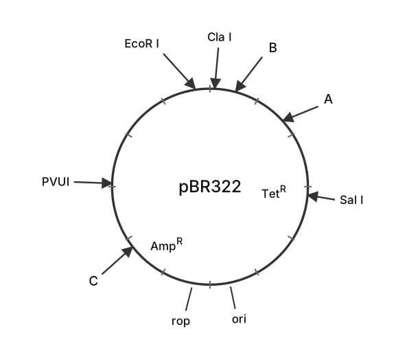
- AP EAPCET 2024Set ap-2024-05-17-AN1 markMCQQ.[FIGURE] (a circular restriction map of plasmid PBR322: sites EcoRI and ClaI are shown close together near a site labelled B; a site labelled A is shown near the start of the TetR gene, with SalI further along within TetR; a site labelled C is shown near PVUI, adjacent to the AmpR gene; the ori and rop genes are also marked on the circle) Identify A, B and C in the diagram of PBR322 (A, B and C are restriction sites) (A) A - BamH I, B - Hind III, C - Pst II (B) A - BamH I, B - Hind III, C - Pst I (C) A - Hind III, B - BamH I, C - Pst II (D) A - Pst I, B - PVU II, C - BamH I
›Reveal solutionSolution
The key is to recall the standard pBR322 restriction map: the unique sites near the TetR gene are BamHI and HindIII, and the site near AmpR is PstI. Matching the figure’s labels gives A = BamHI, B = HindIII, C = PstI, so the correct choice is (B).
The question asks you to identify three unlabeled restriction sites (A, B, C) on the classic plasmid pBR322. This plasmid is one of the most well‑known cloning vectors, and its restriction map is a standard reference in molecular biology. The figure shows the relative order of several known sites (EcoRI, ClaI, SalI, PVUI) and three unknown ones. To solve this, you need to recall the actual map of pBR322 and match the positions.
Why this approach works:
Instead of memorizing every possible combination, focus on the landmark genes (TetR and AmpR) and the unique restriction sites that are commonly used for cloning. In pBR322, the tetracycline‑resistance gene (TetR) contains unique sites for BamHI and HindIII near its start, and the ampicillin‑resistance gene (AmpR) contains a unique PstI site. The figure places site A near the start of TetR, site B near the top (close to EcoRI and ClaI), and site C next to AmpR. By matching these positions to the known map, you can deduce the identities.
Let’s work through it step by step.
-
Recall the standard pBR322 restriction map.
The plasmid is 4361 base pairs long. The key features (clockwise from the EcoRI site at position 0/4361) are:
- EcoRI (0)
- ClaI (23)
- HindIII (29)
- BamHI (375)
- SalI (651)
- TetR gene (positions ~86–1276)
- ori (origin of replication, ~2535)
- rop (repressor of primer, ~1915–2100)
- AmpR gene (positions ~3293–4153)
- PstI (3609) — inside AmpR
- PVUI (3735) — also inside AmpR, near PstI
Notice that HindIII and BamHI are both in the TetR region, but HindIII is much closer to EcoRI/ClaI (at position 29) while BamHI is farther downstream (position 375). PstI is inside the AmpR gene, near PVUI.
-
Match the figure’s labels to these positions.
The figure shows (clockwise from top):
- EcoRI and ClaI (close together at the top)
- B (just to the right of ClaI, still near the top)
- A (further clockwise, near the start of TetR)
- SalI (further along, within TetR)
- TetR (inner label)
- ori and rop (bottom)
- AmpR (inner label, lower left)
- C (next to AmpR)
- PVUI (left side, near C)
From the map above, the site immediately after ClaI (position 23) is HindIII (position 29). So B must be HindIII.
The site near the start of TetR, but before SalI (position 651), is BamHI (position 375). So A must be BamHI.
The site next to AmpR and near PVUI (position 3735) is PstI (position 3609). So C must be PstI.
-
Check the answer choices.
- (A) A‑BamHI, B‑HindIII, C‑PstII → PstII is not a standard pBR322 site; PstI is correct.
- (B) A‑BamHI, B‑HindIII, C‑PstI → matches our deduction.
- (C) A‑HindIII, B‑BamHI, C‑PstII → swaps A and B, and uses PstII.
- (D) A‑PstI, B‑PVUII, C‑BamHI → completely wrong order.
Only option (B) fits the known map.
TipA common pitfall is confusing the order of HindIII and BamHI. Remember: HindIII is very close to EcoRI/ClaI (position 29), while BamHI is farther into the TetR gene (position 375). In the figure, site B is right next to ClaI, so it must be HindIII.
Watch outSome students misread the figure and think site A is at the top near EcoRI, but the arrow for A is clearly placed further clockwise, near the TetR label. Always trace the clockwise order from the known EcoRI site.
✓Final answerThe correct option is (B).
ANSWER: B
-
- AP EAPCET 2023Set ap-2023-05-23-AN1 markMCQQ.In EcoRI, R and I denote (A) Remove nucleotides and cut with in DNA. (B) Name of strain and order of enzyme isolated from the strain of bacteria. (C) Restriction endonuclease and number of strain. (D) Name of the scientist and number of bacteria.
›Reveal solutionSolution
Restriction enzyme names follow a convention: genus+species initials, then strain, then the order of discovery in that strain. In EcoRI, 'R' = strain identifier and 'I' = order of isolation — option (B).
Concept and Intuition
The nomenclature of restriction endonucleases (proposed by Smith and Nathans) encodes real biological information about where the enzyme came from. The first letter (capitalised) is from the genus, the next two letters from the species, giving a three-letter abbreviation (e.g., 'Eco' for Escherichia coli). Any further letter denotes the particular strain of that organism the enzyme was isolated from, and a Roman numeral at the end denotes the order in which that enzyme was identified when a strain produces more than one restriction enzyme.
Step-by-Step Solution
- Break down 'EcoRI': E = genus Escherichia, co = species coli.
- The next letter, R, denotes the specific strain of E. coli (strain RY13) from which this enzyme was isolated.
- The Roman numeral I denotes that this was the first restriction enzyme isolated/identified from that strain.
- Matching this to the options, (B) correctly states 'Name of strain and order of enzyme isolated from the strain of bacteria.'
Common Mistakes
- Confusing 'R' with 'Restriction' — R actually denotes the bacterial strain, not the word "restriction" itself.
- Thinking the Roman numeral refers to the bacterium's serial number rather than the enzyme's order of discovery.
✓Final answerThe correct option is (B) — Name of strain and order of enzyme isolated from the strain of bacteria.
ANSWER: B
- AP EAPCET 2023Set ap-2023-05-23-AN1 markMCQQ.Choose the correct statements. A. Asexual reproduction preserves genetic information. B. Hybridization lead to multiply desirable genes. C. Restriction enzyme adds methyl group to DNA. D. Genetic engineering changes the phenotype of the organism. (A) A and D (B) B and C (C) C and D (D) A and C
›Reveal solutionSolution
Statement A (asexual reproduction preserves genotype) and D (genetic engineering changes phenotype) are correct; B misdescribes hybridisation and C wrongly attributes DNA methylation to restriction enzymes.
Concept and Intuition
Asexual reproduction (mitotic, no gamete fusion) produces offspring genetically identical to the parent — no recombination occurs, so genetic information is preserved unchanged across generations (this is the basis of cloning). Genetic engineering deliberately alters an organism's genome by inserting a foreign gene, and since genotype determines phenotype, this changes the phenotype (e.g., Bt cotton, insulin-producing bacteria). By contrast, a restriction endonuclease's job is to cut DNA at a specific recognition sequence; adding a methyl group to protect a bacterium's own DNA from its own restriction enzyme is the job of a separate modification (methylase) enzyme, not the restriction enzyme itself. And classical hybridisation (crossing different genotypes/varieties) recombines/combines existing desirable traits from two parents — it does not 'multiply' genes in the sense of increasing their copy number.
Step-by-Step Solution
- Evaluate A: asexual reproduction gives genetically identical progeny → preserves genetic information → True.
- Evaluate B: hybridisation combines traits from two genotypes but does not literally 'multiply' desirable genes → False as worded.
- Evaluate C: methylation of DNA is done by a modification enzyme (methylase), not the restriction endonuclease → False.
- Evaluate D: introducing new genes via genetic engineering alters the organism's traits, i.e., its phenotype → True.
- The correct statements are A and D, matching option (A).
Common Mistakes
- Assuming restriction enzymes both cut AND methylate DNA — these are two distinct enzyme classes in the host restriction-modification system.
- Reading 'hybridisation' loosely as synonymous with genetic engineering's ability to combine/introduce genes at will.
✓Final answerThe correct option is (A) — A and D.
ANSWER: A
- AP EAPCET 2023Set ap-2023-05-23-FN1 markMCQQ.5′−GAATTC−3′ 3′−CTTAAG−5′ Mention the Nitrogen base sequence name, restriction cut sites and type of cut ends in the above DNA fragment (A) Recognition sequence, A & A, cohesive ends (B) Palindrome sequence, G & A, sticky ends (C) Endonuclease, T & T, staggered cut (D) Ligase sites, C & T, sticky ends
›Reveal solutionSolution
Recognising the EcoRI restriction site: a palindromic hexanucleotide cut between G and A on each strand, producing sticky ends.
Concept and Intuition
5′−GAATTC−3′ paired with 3′−CTTAAG−5′ reads the same sequence (GAATTC) in the 5′→3′ direction on both strands — the defining property of a palindromic sequence, which is what most restriction enzymes recognise. This particular sequence is the recognition site of the enzyme EcoRI, which cuts between the G and the A on each strand (GAATTC), leaving short single-stranded overhangs at each end — called sticky (cohesive) ends — that can base-pair with complementary overhangs from other DNA cut by the same enzyme.
Step-by-Step Solution
- Check the sequence's directional symmetry: reading 5'→3' on the top strand gives GAATTC; reading 5'→3' on the bottom strand also gives GAATTC — a palindrome.
- Identify the cut position: restriction enzymes recognising this site (EcoRI) cut between the G and A on each strand.
- Because the two cuts are staggered (not directly opposite each other), the resulting fragment ends have short single-stranded overhangs, called sticky or cohesive ends.
- This matches option (B): Palindrome sequence, G & A, sticky ends.
Common Mistakes
- Calling this sequence a "recognition sequence" without noting its more specific defining property of being palindromic (which is what makes recognition sequences of restriction enzymes special).
✓Final answerThe correct option is (B) — Palindrome sequence, G & A, sticky ends.
ANSWER: B
- AP EAPCET 2022Set ap-2022-07-12-AN1 markMCQQ.Two enzymes responsible for restricting the growth of bacteriophage were isolated from (A) Salmonella (B) Agro bacterium (C) Escherichia Coli (D) Archea bacteria
›Reveal solutionSolution
Restriction enzymes were discovered in studies of Escherichia coli, where bacteria used these enzymes to cut and destroy invading bacteriophage DNA, thereby restricting phage growth.
Concept and Intuition
Restriction enzymes (restriction endonucleases) are bacterial enzymes that recognize specific short DNA sequences and cut the DNA at or near those sites. They form part of a bacterium's natural defence system against bacteriophages: while a bacterium methylates and protects its own DNA at these recognition sites, unmethylated foreign (phage) DNA entering the cell gets cleaved, restricting the phage's ability to replicate. The phenomenon and the first restriction enzymes were discovered and characterized in studies on E. coli strains, where researchers noticed that phages grown on one E. coli strain were "restricted" (failed to infect efficiently) when tried on a different E. coli strain — leading to the identification of the responsible restriction-modification enzyme system.
Step-by-Step Solution
- Recall the historical context: restriction enzymes were discovered through host-specific restriction of bacteriophage growth.
- Recall this phenomenon and its causative enzymes were first characterized in Escherichia coli.
- Eliminate Salmonella, Agrobacterium, and Archaea as not being the organism associated with this discovery.
- Select Escherichia coli as the source organism.
Common Mistakes
- Confusing the SOURCE organism of the classic restriction enzymes (E. coli) with Agrobacterium, which is instead associated with the Ti plasmid and plant transformation, not restriction enzyme discovery.
✓Final answerThe correct option is (C) — Escherichia Coli.
ANSWER: C
- AP EAPCET 2022Set ap-2022-07-12-AN1 markMCQQ.Restriction enzyme Digestion means (A) Cutting of DNA (B) Repair of DNA (C) Incubating of DNA (D) Enzymes digestion of DNA
›Reveal solutionSolution
Restriction digestion simply means using restriction enzymes to cut DNA at their specific recognition sequences, generating defined fragments for cloning.
Concept and Intuition
In genetic engineering, DNA needs to be cut precisely so that a gene of interest can be isolated and later ligated into a vector. Restriction endonucleases achieve this by recognizing a specific palindromic DNA sequence and cleaving the phosphodiester backbone at (or near) that site, generating either blunt ends or sticky (cohesive) ends with short single-stranded overhangs. This entire step — treating a DNA sample with a restriction enzyme so it is cleaved at its recognition sites — is what is meant by "restriction digestion." It is purely a cutting process, not repair, incubation, or generic "enzyme digestion" in a vague sense.
Step-by-Step Solution
- Recall what a restriction endonuclease does: recognizes a specific sequence and cuts the DNA there.
- "Restriction digestion" is the standard lab term for subjecting a DNA sample to this enzymatic cutting.
- Rule out "repair of DNA" — that is the role of DNA repair enzymes/ligase, not restriction enzymes.
- Rule out vague/incorrect distractors like "incubating DNA" (a step in the protocol, not the defining action) or overly generic "enzyme digestion of DNA" without specifying cutting.
- Select "cutting of DNA" as the precise meaning of restriction digestion.
Common Mistakes
- Confusing "digestion" (cutting) with "ligation" (joining) — these are opposite steps in cloning.
- Treating "incubating DNA" (a procedural condition needed for the enzyme to act) as the definition itself.
✓Final answerThe correct option is (A) — Cutting of DNA.
ANSWER: A
🎓Unlock everything free for 14 days
- ✓Full step-by-step solutions
- ✓Concept-first explanations
- ✓Methods, shortcuts & mistakes
- ✓PYQ mapping + timed mock tests
Full access for 14 days. No credit card required.