Q.(A) Define the following term :
🔒You're viewing a preview — the full solution, concept, methods & PYQ mapping are locked.
🔒 Start your 14-day free trial to unlock the full solution →Part (a)Concept understanding — Protein Structure Levels
Protein Structure Levels: From a String to a Working Machine
Imagine you have a long string of beads. Each bead is a different colour, and the order of colours is fixed. If you just lay that string on a table, it's a floppy, useless line. But if you could somehow make that string fold itself into a tiny, precise 3D shape — say, a key that fits a specific lock — you'd have something that actually does a job. That's exactly what a protein is.
A protein starts as a long chain of smaller units called amino acids. There are 20 different kinds, each with a unique side chain (the "colour" of the bead). The exact sequence of these amino acids is determined by your DNA. But a protein isn't just a chain — it's a chain that folds into a specific shape, and that shape determines what the protein does. If the shape is wrong, the protein can't work.
The folding happens in stages, and we call these stages the four levels of protein structure.
Level 1: Primary Structure — The Sequence
This is the simplest level: just the linear order of amino acids in the chain, linked by peptide bonds. Think of it as the sentence written in the language of proteins.
Primary structure = the sequence of amino acids from the N-terminus (start) to the C-terminus (end).
Why does this matter? Because the sequence determines everything else. Change one amino acid in a critical spot, and the entire protein can misfold. Example: sickle cell anaemia is caused by a single amino acid swap in haemoglobin — valine replaces glutamic acid at position 6. One bead out of hundreds changes colour, and the whole protein folds wrong.
Level 2: Secondary Structure — Local Folding Patterns
The chain doesn't stay straight. Hydrogen bonds form between the backbone atoms (not the side chains) of nearby amino acids. These bonds cause the chain to twist or fold into regular, repeating patterns.
Two common patterns:
- Alpha helix (α-helix): The chain coils like a spring or a spiral staircase. Hydrogen bonds form between every 4th amino acid, holding the coil tight.
- Beta sheet (β-sheet): The chain folds back and forth like a pleated fan. Hydrogen bonds form between adjacent segments, creating a flat, sheet-like structure.
Secondary structure is stabilised entirely by hydrogen bonds between the carbonyl oxygen of one amino acid and the amide hydrogen of another — both part of the peptide backbone. Side chains stick out and don't participate.
These patterns are local — they happen in short stretches of the chain. A single protein can have multiple α-helices and β-sheets separated by loops.
Level 3: Tertiary Structure — The Global 3D Shape
Now the whole chain folds into its final, compact, three-dimensional shape. This is where the protein becomes functional. The tertiary structure is stabilised by interactions between the side chains of amino acids that may be far apart in the sequence but come close in space.
What holds it together?
- Hydrophobic interactions: Nonpolar side chains cluster together in the protein's interior, away from water.
- Hydrogen bonds: Between polar side chains.
- Ionic bonds: Between positively and negatively charged side chains.
- Disulfide bridges: Covalent bonds between the sulfur atoms of two cysteine amino acids — these are strong and lock parts of the chain together.
- Van der Waals forces: Weak attractions between closely packed atoms.
A common mistake: thinking tertiary structure is just "more secondary structure." It's not. Secondary structure is local folding; tertiary structure is the global arrangement of the entire chain, including how helices and sheets pack together.
Level 4: Quaternary Structure — Multiple Chains Working Together
Some proteins are made of more than one polypeptide chain. Each chain is a separate subunit, and the quaternary structure describes how these subunits assemble into a functional complex. …
Why this formula?
Protein Structure Levels: Understanding the "Why" Behind the Hierarchy
Protein structure is not defined by a single formula, but by a logical hierarchy of organization. Each level builds on the previous one, and the "formulas" here are really principles of molecular interaction that explain why proteins fold the way they do.
Let's break down each level and the reasoning behind its key features.
1. Primary Structure: The Sequence "Formula"
What it is: The linear sequence of amino acids linked by peptide bonds.
Key "formula":
Protein=NH2-[Amino Acid]1-[AA]2-...-[AA]n-COOH
Why this holds:
- Peptide bond formation is a condensation reaction:
-COOH+NH2-→-CO-NH-+H2O
- This bond is rigid and planar due to resonance (partial double-bond character). This restricts rotation, which directly influences higher-order folding.
- The sequence is determined by DNA (genetic code). Every change in sequence can alter the entire structure — this is why a single mutation (e.g., sickle cell anemia: Glu → Val at position 6) can cause disease.
Exam insight: The primary structure is the only level that is covalently determined. All higher levels are non-covalent interactions.
2. Secondary Structure: Local Folding Patterns
Key patterns: α-helix and β-pleated sheet.
Why these form — the hydrogen bond "formula":
The α-helix
- Hydrogen bonds form between the carbonyl oxygen (C=O) of residue n and the amide hydrogen (N-H) of residue n+4.
- Why n+4? This spacing allows the backbone to coil into a right-handed helix with exactly 3.6 amino acids per turn.
- Reasoning: The peptide bond's planar nature forces the backbone into a specific geometry. The n+4 pattern maximizes H-bonding while minimizing steric clashes.
The β-sheet
- Hydrogen bonds form between adjacent strands (either parallel or antiparallel).
- Why not n+4? The backbone is extended (pleated), so H-bonds occur between different segments, not within the same chain.
Key formula (Ramachandran plot):
Only certain backbone dihedral angles (ϕ,ψ) are allowed:
- α-helix: ϕ≈−57∘, ψ≈−47∘
- β-sheet: ϕ≈−130∘, ψ≈+130∘
Why these angles? Steric hindrance — atoms cannot overlap. The Ramachandran plot shows the only regions where no two atoms clash.
3. Tertiary Structure: The 3D Fold
Key "formula": The hydrophobic effect drives folding.
Why this holds:
- Water molecules form a cage-like structure around nonpolar (hydrophobic) side chains. This is entropically unfavorable (water loses freedom).
- To minimize this, hydrophobic side chains cluster together in the protein's core, away from water.
- Result: The protein collapses into a compact globule, with polar/charged residues on the surface.
Supporting interactions (the "glue"):
| Interaction | Why it matters |
|---|---|
| Hydrogen bonds | Between side chains (e.g., Ser–Glu) |
| Ionic bonds | Between charged groups (e.g., Lys–Asp) |
| Van der Waals forces | Close packing of atoms |
| Disulfide bridges | Covalent S–S bonds (only in oxidizing environments) |
Why not just one formula? Tertiary structure is unique to each protein — it's the sum of all these interactions, not a single equation.
4. Quaternary Structure: Multiple Subunits
Key "formula":
Functional protein=∑i=1nSubuniti
Why this holds:
- Some proteins need multiple polypeptide chains to function (e.g., hemoglobin: α2β2). …
Part (b)Concept understanding — Glucose Cyclization
Glucose Cyclization: From a Straight Chain to a Ring
Imagine you have a long, flexible chain with a hook at one end and an eyelet at the other. If you swing that chain around, the hook can snap into the eyelet, forming a loop. That's the core idea behind glucose cyclization.
Glucose in its simplest written form is a straight chain of six carbon atoms with an aldehyde group (−CHO) at one end. But in water (like in your blood), this chain doesn't stay straight. The aldehyde group reacts with the alcohol group on the fifth carbon, forming a stable six-membered ring.
Why does this happen?
The aldehyde carbon is electrophilic (electron-poor), and the oxygen on carbon-5 has lone pairs (nucleophilic). They attack each other, forming a new bond. This creates a hemiacetal — a carbon bonded to both an −OH and an −OR group. The ring is more stable than the open chain in solution.
The open-chain form of glucose exists in equilibrium with the cyclic form, but at any given time, over 99% of glucose molecules are in the cyclic form.
The precise statement
Glucose cyclization is an intramolecular nucleophilic addition where the aldehyde group at C1 reacts with the hydroxyl group at C5, forming a six-membered pyranose ring (named after pyran, a six-membered oxygen heterocycle). This reaction creates a new chiral center at C1, giving two possible stereoisomers called anomers: α and β.
The two anomers
When the ring forms, the oxygen from C5 becomes part of the ring. The carbon that was the aldehyde (now C1) becomes a new chiral center. The −OH group at this new center can point:
- Down (relative to the ring plane) → α-D-glucose
- Up → β-D-glucose
The α and β anomers are diastereomers, not enantiomers. They differ only at the anomeric carbon (C1). In solution, they interconvert through the open-chain form — a process called mutarotation.
How to draw it (Haworth projection)
- Draw a hexagon with an oxygen atom at the top-right corner.
- Number the carbons clockwise from the oxygen: C1 is the carbon to the right of oxygen, C2 next, and so on.
- For α-D-glucose, the −OH at C1 points down (opposite to the CH2OH group at C5).
- For β-D-glucose, the −OH at C1 points up (same side as the CH2OH group). …
Why this formula?
Glucose Cyclization: Why the Ring Forms
Glucose cyclization is a classic example of an intramolecular reaction — a molecule reacting with itself. Let's build the understanding step by step.
1. The Starting Point: Open-Chain Glucose
Glucose (C6H12O6) in its open-chain form has:
- An aldehyde group (−CHO) at carbon 1 (C1)
- A hydroxyl group (−OH) at carbon 5 (C5)
The aldehyde is electrophilic (electron-deficient at the carbonyl carbon), and the hydroxyl is nucleophilic (electron-rich oxygen with lone pairs).
2. Why Cyclization Happens: Thermodynamic & Kinetic Favorability
The Key Insight
The molecule is flexible — it can bend so that the C5 hydroxyl oxygen approaches the C1 aldehyde carbon. This brings two reactive groups into close proximity.
- Entropy is favorable: One molecule becomes one ring — no loss of translational entropy (unlike two separate molecules reacting).
- Ring strain is manageable: A 5- or 6-membered ring (furanose or pyranose) has minimal angle strain (close to tetrahedral angles).
Result: The reaction is reversible but strongly favors the cyclic form — in aqueous solution, >99% of glucose exists as the ring.
3. The Reaction: Hemiacetal Formation
The nucleophilic oxygen of the C5 hydroxyl attacks the electrophilic carbonyl carbon of C1:
R-CHO+R’-OH⇌R-CH(OH)(OR’)
This is a hemiacetal — a carbon bonded to both an −OH and an −OR group.
Mechanism (simplified)
- Protonation of the carbonyl oxygen (acid-catalyzed) makes C1 more electrophilic.
- Nucleophilic attack by the C5 oxygen.
- Deprotonation yields the cyclic hemiacetal.
4. The Key Formula(e): Ring Size & Anomeric Carbon
Ring Size Determination
The ring size depends on which hydroxyl attacks:
- C5 hydroxyl → pyranose (6-membered ring: 5 carbons + 1 oxygen)
- C4 hydroxyl → furanose (5-membered ring: 4 carbons + 1 oxygen)
For D-glucose, the C5 attack is overwhelmingly favored, giving the pyranose form.
The Anomeric Carbon
The new chiral center formed at C1 is called the anomeric carbon. Two stereoisomers arise:
- α-anomer: −OH at C1 is trans to the −CH2OH group (axial in the chair conformation)
- β-anomer: −OH at C1 is cis to the −CH2OH group (equatorial in the chair conformation)
Why two forms? The attack can occur from either face of the planar carbonyl group — leading to two possible configurations at the new stereocenter.
5. The Equilibrium Constant & Mutarotation
The interconversion between α and β anomers is called mutarotation:
α-D-glucopyranose⇌open chain⇌β-D-glucopyranose
At equilibrium (in water at 20°C):
- β-D-glucopyranose: ~64%
- α-D-glucopyranose: ~36%
- Open chain: <0.1% …
Part (a)
Reducing sugar: a carbohydrate carrying a free aldehyde or keto group (a free anomeric carbon) that can reduce Tollens' or Fehling's reagent. All monosaccharides and most disaccharides (maltose, lactose) are reducing; sucrose is not.
Fibrous vs globular proteins: fibrous are long, thread-like, water-insoluble, structural (keratin, collagen); globular are spherical, water-soluble, functional (enzymes, haemoglobin). …
Part (a): a reducing sugar has a free aldehyde/keto group; fibrous proteins are insoluble structural, globular are soluble functional; a nucleotide is a nucleoside plus a phosphate. Part (b): glucose + hydroxylamine → oxime; + acetic anhydride → pentaacetate; + conc. HNO₃ → saccharic (glucaric) acid.
Part (a)
- Reducing sugar. A reducing sugar is a carbohydrate that acts as a reducing agent because it possesses a free aldehyde or keto group (a free anomeric –OH). It reduces mild oxidants such as Tollens' (Ag+) or Fehling's (Cu2+) reagent. All monosaccharides (glucose, fructose) and most disaccharides (maltose, lactose) are reducing; sucrose is non-reducing because both anomeric carbons are engaged in the glycosidic bond.
- Differences. (i) Fibrous vs globular proteins
| Feature | Fibrous | Globular |
|---|---|---|
| Shape | long, thread-like | spherical, folded |
| Solubility | insoluble in water | soluble in water |
| Function | structural (keratin, collagen, silk) | functional (enzymes, hormones, haemoglobin) |
(ii) Nucleotide vs nucleoside
| Feature | Nucleoside | Nucleotide |
|---|---|---|
| Composition | base + pentose sugar | base + pentose sugar + phosphate |
Showing the 12 most recent of 36 on this concept.
- AP EAPCET 2026Set eng-2026-05-13-AN1 markMCQQ.The number of secondary alcoholic groups present in the end product 'Y' of the given reaction sequence is C6H12O6HCNXH2O ∣ H+Y (A) 4 (B) 3 (C) 5 (D) 6
›Reveal solutionSolution
This tests the Kiliani–Fischer chain-extension sequence (aldose + HCN → cyanohydrin → hydrolysis → one-carbon-longer aldonic acid) and careful counting of secondary alcohol groups in the product. The answer is 5.
Concept and Intuition
Glucose's open-chain form has an aldehyde at C1. Adding HCN to an aldehyde forms a cyanohydrin: the carbonyl carbon becomes a new stereocentre bearing both −OH and −CN, and crucially the −CN carbon becomes a new carbon added to the chain. When that nitrile is hydrolysed (H2O/H+) it becomes a carboxylic acid carbon. So overall, a 6-carbon aldose becomes a 7-carbon acid with one extra −CH(OH)− unit inserted next to the original carbonyl position — this is exactly how sugar chains are lengthened in the Kiliani–Fischer synthesis.
Step-by-Step Solution
- Write glucose's open-chain structure: C1(CHO)−C2(CHOH)−C3(CHOH)−C4(CHOH)−C5(CHOH)−C6(CH2OH).
- HCN addition at the carbonyl (C1): the carbonyl carbon becomes −CH(OH)(CN)−, i.e. a new nitrile carbon is attached to old C1. So X: NC−CH(OH)[oldC1]−CH(OH)[C2]−CH(OH)[C3]−CH(OH)[C4]−CH(OH)[C5]−CH2OH[C6].
- Hydrolysis (H2O/H+) converts the terminal −CN to −COOH: Y: HOOC[new carbon]−CH(OH)[old C1]−CH(OH)[C2]−CH(OH)[C3]−CH(OH)[C4]−CH(OH)[C5]−CH2OH[C6] — a 7-carbon chain. …
- AP EAPCET 2026Set eng-2026-05-15-FN1 markMCQQ.Observe the following reactions I. D-GlucoseNH2OH II. D-Glucose(i) (CH3CO)2O(ii) NH2OH Correct statement regarding the reactions I and II is (A) Oxime is formed in both the reactions I, II (B) Oxime is not formed in both the reactions I, II (C) Oxime is formed in reaction I but oxime is not formed in reaction II (D) Oxime is not formed in reaction I but oxime is formed in reaction II
›Reveal solutionSolution
This is the classic experiment proving glucose's cyclic hemiacetal structure: plain
glucose reacts with hydroxylamine (free −CHO), but glucose pentaacetate does
not (no free −CHO left to react). Answer: (C).
Concept and Intuition
D-Glucose is not fixed as a straight chain — it exists mainly as a cyclic hemiacetal
(pyranose) in equilibrium with a small amount of the open-chain aldehyde form. Reagents
that test for a free carbonyl (like NH2OH, Schiff's reagent, or NaHSO3) react via
this open-chain minority form, so plain glucose behaves as if it has a free −CHO.
If, however, glucose is first acetylated with excess acetic anhydride, every −OH group gets esterified — including the anomeric −OH that would otherwise open up to
expose the aldehyde. The resulting glucose pentaacetate is locked in the cyclic
form with no accessible carbonyl, so it fails to react with hydroxylamine or Schiff's
reagent. This experiment is one of the classical pieces of evidence for the cyclic
(not open-chain) structure of glucose.
Step-by-Step Solution
- Reaction I: D-GlucoseNH2OHoxime — plain glucose's open-chain aldehyde tautomer reacts normally, forming the oxime.
- Reaction II: D-Glucose(i) (CH3CO)2Oglucose pentaacetate — this step acetylates all −OH groups, including the anomeric one, locking the ring closed. …
- AP EAPCET 2026Set ap-2026-05-19-FN1 markMCQQ.Choose the correct statements A) The first amino acid in a protein chain is in the left end and termed as C-terminal amino acids. B) Long protein chain folded upon itself like a hollow woolen ball is called tertiary structure. C) In general, only right handed helices are observed in protein. D) In general, polysaccharides will have glycosidic bond formed by dehydration. (A) A, B and C (B) B, C and D (C) A, C and D (D) A, B and D
›Reveal solutionSolution
Of four claims about protein/polysaccharide structure, only the "first amino acid = C-terminal" claim is wrong (it should be N-terminal); tertiary structure, right-handed helices, and dehydration-formed glycosidic bonds are all correct.
Concept and Intuition
A polypeptide has directionality: one end carries a free –NH₂ group (the N-terminus, conventionally drawn/numbered first, "left" end) and the other a free –COOH group (the C-terminus, "right" end). Beyond primary structure (sequence) and secondary structure (regular local folding like the α-helix, predominantly right-handed in natural proteins), further compact 3-D folding into a globular shape is tertiary structure. Separately, polysaccharides are built by joining monosaccharide units via glycosidic bonds, formed through dehydration (loss of a water molecule per bond), just like peptide bonds in proteins.
Step-by-Step Solution
- A: the residue with the free amino group is by convention the FIRST/N-terminal amino acid (drawn on the left) — calling it "C-terminal" is a direct mix-up of the two termini. FALSE.
- B: further folding of an already-folded (secondary structure) polypeptide chain into a compact, roughly spherical, hollow-ball-like shape is precisely the description used for tertiary structure. TRUE. …
- AP EAPCET 2026Set ap-2026-05-20-AN1 markMCQQ.D-Glucose does not react with which of the following reagents? I. NaHSO3 II. NH2OH III. (CH3CO)2O IV. Schiff's reagent (A) II, III only (B) I, II, III only (C) I, II only (D) I, IV only
›Reveal solutionSolution
Glucose fails to react with sodium bisulphite and Schiff's reagent — the classic evidence that its carbonyl exists mainly as a cyclic hemiacetal, not a free aldehyde.
Concept and Intuition
Glucose behaves like a typical aldehyde in many respects (oxidation to gluconic acid, cyanohydrin formation, oxime formation, acetylation of its five -OH groups), which is used to establish its structure. But it notably FAILS the two classic 'free aldehyde' spot tests — the bisulphite addition reaction and the Schiff's test — because in solution glucose exists overwhelmingly in the cyclic hemiacetal (pyranose) form, and the small equilibrium amount of open-chain aldehyde isn't sufficient/fast enough for these particular fast, stoichiometric addition reactions, even though it's enough to drive the slower oxidation and condensation reactions via Le Chatelier's principle.
Step-by-Step Solution
- I. NaHSO3: Glucose does NOT form the expected bisulphite addition compound — a key piece of evidence against a simple free-aldehyde structure.
- II. NH2OH: Glucose DOES react, forming an oxime, confirming the presence of a carbonyl group (via the open-chain tautomer).
- III. (CH3CO)2O: Glucose DOES react, undergoing acetylation to give glucose pentaacetate (confirms 5 -OH groups). …
- AP EAPCET 2025Set eng-2025-05-21-AN1 markMCQQ.A carbonyl compound X(C8H8O) gives yellow precipitate with NaOI. Hemiacetal of X with methanol/dry HCl is (A) C6H5−CH2−CH(OH)−OCH3 (drawn as a benzene ring with a −CH2CH(OH)OCH3 substituent) (B) C6H5−CH2−CH(OCH3)2 (drawn as a benzene ring with a −CH2CH(OCH3)2 substituent) (C) C6H5−C(OH)(CH3)(OCH3) (drawn as a benzene ring whose ring carbon bears a central carbon substituted with -OH, -CH_3 and -OCH_3) (D) C6H5−C(OH)(OCH3)2 (drawn as a benzene ring whose ring carbon bears a central carbon substituted with -OH and two -OCH_3 groups)
›Reveal solutionSolution
X is acetophenone (C6H5COCH3, iodoform-positive, C8H8O); its hemiacetal with methanol is C6H5−C(OH)(CH3)(OCH3).
Concept and Intuition
The iodoform (yellow precipitate with NaOI, i.e. I2/NaOH) test is positive for any compound with a CH3−CO− group (methyl ketones) or that can be oxidised to one (like ethanol/secondary alcohols bearing a CH3CH(OH)− group). Given the molecular formula C8H8O and an aromatic-sized carbon count, the methyl ketone that fits is acetophenone, C6H5−CO−CH3. A hemiacetal forms when one equivalent of alcohol adds across a carbonyl (catalysed by dry HCl): the carbonyl oxygen picks up the proton to become −OH, while the alcohol's oxygen bonds to the former carbonyl carbon as −OR — all substituents originally on that carbon (here, the phenyl and the methyl) remain attached to the same, now sp3, carbon.
Step-by-Step Solution
- Identify X: C8H8O, gives iodoform test ⇒ a methyl ketone. Acetophenone C6H5COCH3 fits (6 (ring)+1 (C=O)+1 (CH3)=8 carbons; one O).
- Hemiacetal formation mechanism: methanol's OH oxygen attacks the electrophilic carbonyl carbon of C6H5−C(=O)−CH3, protonated by dry HCl. …
- AP EAPCET 2025Set eng-2025-05-22-FN1 markMCQQ.Which of the following contain α-D-glucose units ?a) cane sugar b) milk sugar c) cellulose d) amylose (A) a, d (B) a, b (C) b, c (D) c, d
›Reveal solutionSolution
Tests recognising which common carbohydrates are actually built from α-D-glucose units, as opposed to β-D-glucose or a non-glucose sugar.
Concept and Intuition
A saccharide "contains α-D-glucose units" only when a glucose unit's anomeric carbon is tied down in the α configuration by a glycosidic bond (as in a polysaccharide chain, or in sucrose where glucose's anomeric carbon links to fructose), or the free sugar is drawn/crystallised as its α anomer feeding a fixed linkage.
Step-by-Step Solution
- (a) Cane sugar = sucrose = α-D-glucose (1→2) β-D-fructose. The glucose unit's anomeric carbon (C1) is locked in the α configuration by this glycosidic bond — contains α-D-glucose. ✓
- (b) Milk sugar = lactose = β-D-galactose (1→4) D-glucose. Here it is galactose, not glucose, that supplies its anomeric carbon to the bond; the glucose unit's own anomeric carbon is FREE (this is why lactose is a reducing sugar and shows mutarotation) — not fixed as α-D-glucose. ✗
- (c) Cellulose is a linear polymer of β-D-glucose units joined by β(1→4) linkages — glucose, but the WRONG anomer (β, not α). ✗ …
- AP EAPCET 2025Set eng-2025-05-23-FN1 markMCQQ.Consider the following. Statement-I : Primary structure of protein represents its constitution Statement-II : α-Helix and β-pleated sheet structure of protein represent tertiary structure of it Correct answer is (A) Both statement-I and statement-II are correct (B) Both statement-I and statement-II are not correct (C) Statement-I is correct, but statement-II is not correct (D) Statement-I is not correct, but statement-II is correct
›Reveal solutionSolution
This tests the protein structure hierarchy: primary = sequence (constitution), secondary = local folding patterns like α-helix/β-sheet, tertiary = overall 3-D folding. Statement-I is right; Statement-II wrongly calls a secondary-structure feature 'tertiary'.
Concept and Intuition
Proteins are described at four structural levels: primary (linear amino-acid sequence — the actual covalent 'constitution' of the molecule), secondary (regular, repeating local folding patterns held by hydrogen bonds along the peptide backbone — the α-helix and β-pleated sheet are the two classic examples), tertiary (the overall three-dimensional folding of the whole chain, stabilised by disulfide bonds, hydrogen bonds, and hydrophobic interactions among side chains), and quaternary (arrangement of multiple polypeptide subunits). Recognising which level a named feature belongs to is the entire test here.
Step-by-Step Solution
- Statement-I: the primary structure is defined as the sequence in which amino acids are linked via peptide bonds — this is indeed the molecule's basic constitution (which amino acid, in which order). True. …
- AP EAPCET 2025Set ap-2025-05-19-AN1 markMCQQ.During the denaturation of proteins, which of the following structures will remain intact? (A) Secondary & tertiary (2∘, 3∘) (B) Tertiary only (3∘) (C) Primary only (1∘) (D) Primary and secondary (1∘, 2∘)
›Reveal solutionSolution
Denaturation breaks the weak interactions maintaining secondary/tertiary folding, but the covalent peptide-bond backbone (primary structure) survives untouched.
Concept and Intuition
Protein structure is organised in levels: primary (amino-acid sequence, held by strong covalent peptide bonds), secondary (α-helices/β-sheets, held by hydrogen bonds), and tertiary (overall 3-D fold, held by a mix of hydrogen bonds, hydrophobic effects, ionic interactions, and disulfide bridges). Denaturing agents (heat, extreme pH, chaotropic agents) disrupt these weak non-covalent interactions but do not have enough energy to break the strong covalent peptide bonds of the backbone.
Step-by-Step Solution
- Denaturation unfolds the protein by breaking the hydrogen bonds/hydrophobic interactions responsible for secondary structure.
- It also destroys tertiary structure (loss of the specific 3-D globular shape), since tertiary structure depends on similarly weak interactions (plus occasionally disulfide bonds, which some strong denaturants can also break). …
- AP EAPCET 2025Set ap-2025-05-19-FN1 markMCQQ.Match the following List-I A) Glycosidic bond B) Hollow woollen ball folding C) Enzyme D) Esterbond List-II I) Trypsin II) Bond between phosphate and 5th carbon of sugar III) tertiary structure of protein IV) formed by dehydration (A) A-II, B-I, C-IV, D-III (B) A-IV, B-III, C-I, D-II (C) A-III, B-II, C-I, D-IV (D) A-IV, B-I, C-II, D-III
›Reveal solutionSolution
Matching each biomolecule term to its correct description gives A-IV, B-III, C-I, D-II — answer (B).
Concept and Intuition
- Glycosidic bond: forms between two monosaccharides via a dehydration (condensation) reaction, releasing a water molecule.
- The image of a protein folding "like a hollow woollen ball" is a common descriptive analogy for a protein's tertiary structure — the overall 3D folding of a single polypeptide chain.
- Trypsin is a well-known digestive protease and is the standard textbook example cited for "enzyme."
- The ester bond joining a phosphate group to the 5′-carbon hydroxyl of a sugar (as in nucleotides) is a phosphoester linkage.
Step-by-Step Solution
- A) Glycosidic bond → IV (formed by dehydration).
- B) "Hollow woollen ball" folding → III (tertiary structure of protein).
- C) Enzyme → I (Trypsin, a textbook enzyme example). …
- AP EAPCET 2025Set ap-2025-05-19-FN1 markMCQQ.Choose the correct statements among the following I. In exothermic reactions 'P' is at a lower level than 'S'. II. Enzyme activity can be affected by temperature etc. with alteration of tertiary structure of protein. III. Low temperature denature the protein. IV. Ligases can remove the groups from substrate (A) I, II (B) III, IV (C) II, IV (D) I, III
›Reveal solutionSolution
Exothermic reactions do place products at lower energy than substrates, and temperature does affect activity via tertiary-structure changes — both true. Low temperature inactivates (not denatures) enzymes, and ligases join (not remove) — both false. Correct set: I, II — option (A).
Concept and Intuition
Enzyme-catalysed reaction energetics: in an exothermic reaction, the products (P) are at a lower free-energy level than the substrates (S), with the difference released as heat — this is basic thermodynamics of catalysed reactions and is independent of the enzyme itself (the enzyme only lowers the activation energy, not the net energy change). Enzyme activity is highly temperature-sensitive because temperature affects the enzyme's three-dimensional (tertiary) structure, which determines the shape of its active site; this is why activity rises with temperature up to an optimum and then falls off as heat begins to disrupt this structure (denaturation) at high temperatures. Crucially, LOW temperature does not denature an enzyme — it merely reduces the kinetic energy of molecules, slowing the reaction reversibly (activity resumes when temperature rises again); denaturation (irreversible loss of structure) is a HIGH-temperature (or extreme pH) phenomenon. Ligases are enzymes that join two molecules together (often using ATP, e.g., DNA ligase sealing nicks) — they do not remove groups from a substrate; that role belongs to hydrolases/lyases.
Step-by-Step Solution
- Statement I: In exothermic reactions, product (P) is at a lower energy level than substrate (S) — TRUE, by definition of an exothermic process (energy released, product lower in energy). …
- AP EAPCET 2025Set ap-2025-05-20-AN1 markMCQQ.Sucrose when boiled with dilute HCl gives two functional isomers X and Y. X gives monocarboxylic acid with bromine water but not Y. The number of –OH groups in cyclic structure of X is (A) 6 (B) 4 (C) 5 (D) 3
›Reveal solutionSolution
X (from sucrose hydrolysis) is glucose, identified because bromine water oxidises only aldoses; its cyclic pyranose form carries 5 free −OH groups.
Concept and Intuition
Sucrose is a non-reducing disaccharide of glucose and fructose joined through their anomeric carbons. Acid hydrolysis (dil. HCl, heat) breaks the glycosidic bond, releasing free glucose and fructose — this equimolar mixture is called invert sugar. Bromine water is a mild, selective oxidant: it oxidises the free aldehyde group of an aldose (like glucose) to a carboxylic acid, but it does not oxidise ketoses (like fructose), because ketones are far more resistant to this kind of mild oxidation than aldehydes.
Step-by-Step Solution
- Sucrose dil. HCl,Δ glucose + fructose (invert sugar) — these are the two isomeric monosaccharides X and Y (C6H12O6 each).
- Bromine water oxidises the aldehyde of glucose to gluconic acid (a monocarboxylic acid), but does not react with fructose's ketone group. So X = glucose, Y = fructose.
- Draw the cyclic (pyranose, 6-membered) ring of glucose: ring atoms are the ring-O, C1, C2, C3, C4, C5, with C6 (CH2OH) exocyclic on C5. …
- AP EAPCET 2025Set eng-2025-05-27-FN1 markMCQQ.Which of the following represents the correct structure of β−D−(−)-Fructofuranose ? (A) [FIGURE] (Haworth furanose ring with O at the top vertex; upper-left ring carbon carries an exocyclic HOH2C− group, with H outside and H just inside it; upper-right ring carbon carries an exocyclic −CH2OH group, with HO inside and OH outside; bottom-left ring carbon carries OH outside; bottom-right ring carbon carries H outside) (B) [FIGURE] (Haworth furanose ring with O at the top vertex; upper-left ring carbon carries an exocyclic HOH2C− group, with H outside and H inside; upper-right ring carbon carries OH directly, with H inside and CH2OH outside; bottom-left ring carbon carries OH outside; bottom-right ring carbon carries OH outside) (C) [FIGURE] (Haworth furanose ring with O at the top vertex; upper-left ring carbon carries an exocyclic HOH2C− group, with H outside and H inside; upper-right ring carbon carries OH directly, with HO inside and CH2OH outside; bottom-left ring carbon carries OH outside; bottom-right ring carbon carries H outside) (D) [FIGURE] (Haworth furanose ring with O at the top vertex; upper-left ring carbon carries H directly, with H inside; upper-right ring carbon carries OH directly, with HO inside; the exocyclic HOH2C− and −CH2OH groups instead sit on the bottom-left and bottom-right ring carbons respectively, which also carry OH outside and H outside)
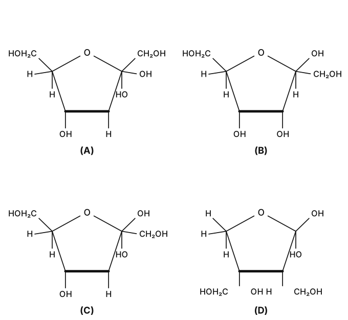
›Reveal solutionSolution
This tests the Haworth structure of β-D-fructofuranose — the 5-membered ring form of D-fructose found in sucrose. Matching each option's substituent placement to the known structure gives option (C).
Concept and Intuition
D-Fructose is a ketohexose; its furanose (5-membered) ring forms between the carbonyl carbon C2 and the C5-oxygen, releasing C1 (CH2OH) and C6 (CH2OH) as exocyclic groups on C2 and C5 respectively. The anomeric carbon here is C2 (not C1, since fructose's carbonyl is a ketone at C2). The α/β distinction refers to the orientation of the new OH generated at C2 upon ring closure, relative to the reference (C5) configuration — exactly analogous to the α/β distinction at C1 in glucopyranose.
Step-by-Step Solution
- Identify the ring: 5-membered furanose ring = O, C2, C3, C4, C5 (C1 and C6 are exocyclic CH2OH groups).
- C5 (derived from a D-sugar's terminal configuration) carries its CH2OH (C6) group "up" in the standard Haworth convention for D-sugars — matching the exocyclic CH2OH shown at the upper-right ring carbon in the options.
- C2 (anomeric) carries the exocyclic HOH2C− (C1) group at the upper-left ring carbon, plus its anomeric OH; for the β-anomer this OH sits on the same face as the C5→C6 reference substituent. …
🎓Unlock everything free for 14 days
- ✓Full step-by-step solutions
- ✓Concept-first explanations
- ✓Methods, shortcuts & mistakes
- ✓PYQ mapping + timed mock tests
Full access for 14 days. No credit card required.