Biology · Ch 11 — Biotechnology: Principles and Processes
Restriction Enzymes
Restriction Enzymes
Restriction enzymes are the molecular scissors of genetic engineering. Their discovery in the 1960s made it possible to cut DNA at precise locations, which is the first step in creating recombinant DNA. Without them, we could not isolate a gene from one organism and insert it into another.
In 1963, two enzymes were isolated from Escherichia coli that together restricted the growth of a bacteriophage (a virus that infects bacteria). One enzyme added methyl groups to the bacterial DNA, protecting it from being cut. The other enzyme cut the viral DNA. This cutting enzyme was named restriction endonuclease.
Five years later, the first restriction endonuclease to be characterised was Hind II. It was special because its cutting action depended on a specific DNA sequence. Hind II always recognised a particular sequence of six base pairs and cut the DNA at that exact point. This specific sequence is called the recognition sequence for Hind II.
Today, over 900 restriction enzymes have been isolated from more than 230 strains of bacteria. Each enzyme recognises a different recognition sequence.
Naming of Restriction Enzymes
The naming convention follows a clear pattern based on the bacterial source:
- The first letter comes from the genus of the prokaryote.
- The next two letters come from the species.
- A following letter (if any) indicates the strain.
- Roman numerals (I, II, III…) show the order in which the enzymes were isolated from that strain.
EcoRI comes from Escherichia coli strain RY 13.
- E = Escherichia (genus)
- co = coli (species)
- R = RY 13 (strain)
- I = first enzyme isolated from that strain
Types of Nucleases
Restriction enzymes belong to a larger class of enzymes called nucleases, which are of two kinds:
- Exonucleases: Remove nucleotides from the ends of the DNA molecule.
- Endonucleases: Make cuts at specific positions within the DNA molecule. Restriction endonucleases are a special type of endonuclease.
How Restriction Enzymes Work
Each restriction endonuclease functions by ‘inspecting’ the length of a DNA sequence. Once it finds its specific recognition sequence, it binds to the DNA and cuts each of the two strands of the double helix at specific points in their sugar-phosphate backbones.
Palindromic Recognition Sequences
Each restriction endonuclease recognises a specific palindromic nucleotide sequence in the DNA.
A palindrome in language is a word that reads the same forwards and backwards (e.g., “MALAYALAM”). In DNA, a palindrome is a sequence of base pairs that reads the same on both strands when the direction of reading (5' → 3') is kept the same.
The recognition sequence for EcoRI is:
5' —— GAATTC —— 3'
3' —— CTTAAG —— 5'
Notice that the sequence on the top strand (5' → 3') is GAATTC, and the sequence on the bottom strand (5' → 3') is also GAATTC (since CTTAAG read backwards is GAATTC).
Sticky Ends
Restriction enzymes cut the DNA strand a little away from the centre of the palindrome site, but between the same two bases on the opposite strands. This leaves single-stranded overhanging stretches at the ends. These are called sticky ends because they can form hydrogen bonds with their complementary cut counterparts.
This stickiness is crucial — it facilitates the action of the enzyme DNA ligase, which seals the cut ends together.
Role in Recombinant DNA Technology
Restriction endonucleases are used to form recombinant molecules of DNA — molecules composed of DNA from different sources or genomes.
When the same restriction enzyme cuts both the source DNA (containing the gene of interest) and the vector DNA (the carrier molecule), both produce the same kind of sticky ends. These complementary sticky ends can then be joined together (end-to-end) using DNA ligase.
Unless you cut the vector and the source DNA with the same restriction enzyme, the recombinant vector molecule cannot be created.
Separation and Isolation of DNA Fragments
After cutting DNA with restriction enzymes, we get fragments of different sizes. These fragments are separated by a technique called gel electrophoresis.
- DNA fragments are negatively charged molecules. …
Drawn by us to help you understand the concept clearly, and verified to make sure it's accurate. For exams, practice from your textbook's own diagram.
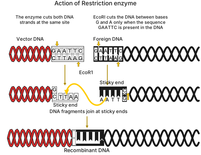
The figure is a 3-step schematic showing what EcoRI does to double-stranded DNA — using labelled arrows and base letters, not a drawn enzyme shape.
Step 1: A stretch of double-stranded DNA is shown with its EcoRI recognition site highlighted — 5-prime-GAATTC-3-prime on one strand, 3-prime-CTTAAG-5-prime on the other. This six-base-pair sequence is a palindrome (it reads the same on both strands in the 5-prime to 3-prime direction), which is what EcoRI recognises.
Step 2: An arrow marks where EcoRI cuts — between the G and the A on each strand. Because the cuts on the two strands are offset, this leaves short, single-stranded overhangs called sticky ends, which are complementary to each other.
Step 3: These sticky ends allow the cut fragment to pair up with any other DNA cut by the same enzyme, and DNA ligase then seals the join, producing a new recombinant strand. …
Drawn by us to help you understand the concept clearly, and verified to make sure it's accurate. For exams, practice from your textbook's own diagram.
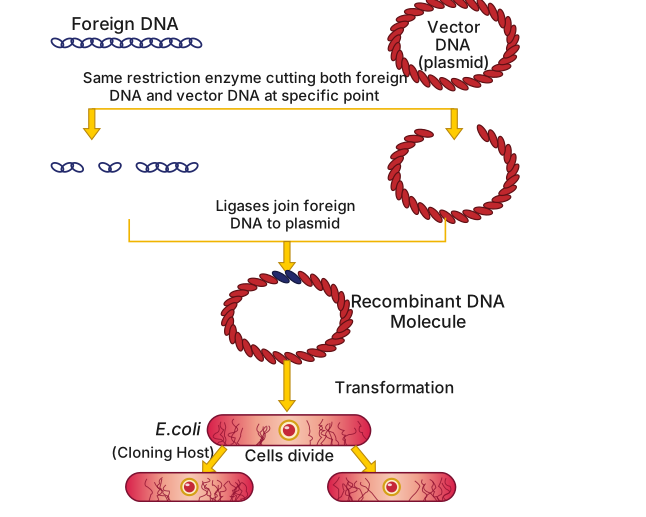
The figure is a numbered-step schematic of the overall recombinant DNA workflow, using plain labelled arrows and text callouts — not scissors or glue-bottle icons.
It starts with two separate DNA molecules: a plasmid cloning vector (small and circular) and the source DNA (containing the gene of interest). Both are cut with the same restriction enzyme, so both end up with matching sticky ends. DNA ligase then joins the gene of interest into the cut vector, producing a recombinant DNA molecule. This recombinant vector is introduced into a host cell, which multiplies — carrying and copying the recombinant DNA along with it. …
Drawn by us to help you understand the concept clearly, and verified to make sure it's accurate. For exams, practice from your textbook's own diagram.
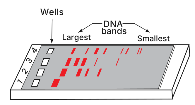
Figure 9.3 is a diagram of an agarose gel as it would appear under UV light after staining with ethidium bromide. The gel is drawn as a 3-D slab viewed at an angle. The loading wells sit at the near/left edge of the slab, in four numbered lanes (1-4). DNA fragments are negatively charged and migrate through the agarose matrix away from the wells, toward the far/right edge of the gel (where the anode, the positive electrode, is positioned off to that side of the tank).
Lane 1 contains undigested DNA. This sample has not been cut by any restriction enzyme, so it remains as a single, large, high-molecular-weight molecule. Because of its size, it cannot move far through the gel matrix; it stays very close to the loading well, appearing as a single bright band close to the well end of the lane.
Lanes 2, 3, and 4 each contain DNA that has been digested with a restriction enzyme. The enzyme has cut the DNA at specific recognition sequences, producing a set of fragments of different lengths. These fragments are now separated by size as they migrate through the agarose gel. The largest fragments move the least and appear as bands closest to the well end of each lane; progressively smaller fragments travel farther across the gel, forming a ladder-like pattern of distinct orange bands. The smallest fragments are found farthest from the wells. …