Biology · Ch 14 — Breathing and Exchange of Gases
Transport of Oxygen
Transport of Oxygen
Oxygen does not travel through the blood on its own; most of it is carried bound to a special pigment inside the red blood cells. That pigment is haemoglobin — a red, iron-containing molecule packed within the RBCs. Because oxygen rides on haemoglobin, the way this pigment picks up and releases oxygen decides how efficiently tissues are supplied.
How oxygen binds
Oxygen attaches to haemoglobin reversibly, forming a compound called oxyhaemoglobin. Reversible means the same bond can form and break depending on the surrounding conditions — haemoglobin grabs oxygen where it is plentiful and lets it go where it is needed. A single haemoglobin molecule can hold at most four oxygen molecules.
Whether oxygen binds depends chiefly on the partial pressure of oxygen — essentially how much oxygen is available in that location. A few other factors also influence the binding:
- the partial pressure of carbon dioxide,
- the hydrogen ion (acidity) concentration, and
- the temperature.
The oxygen dissociation curve
If you plot the percentage saturation of haemoglobin with oxygen against the partial pressure of oxygen, the result is not a straight line but an S-shaped (sigmoid) curve, known as the oxygen dissociation curve. This curve is a useful tool for studying how factors such as carbon dioxide pressure and hydrogen ion concentration affect the loading and unloading of oxygen.
Loading at the lungs, unloading at the tissues
The conditions at the alveoli and at the body tissues are almost opposites, and this is exactly what makes the transport system work:
- In the alveoli — oxygen pressure is high, carbon dioxide pressure is low, hydrogen ion concentration is low, and temperature is lower. Every one of these favours the formation of oxyhaemoglobin, so haemoglobin picks up oxygen here. …
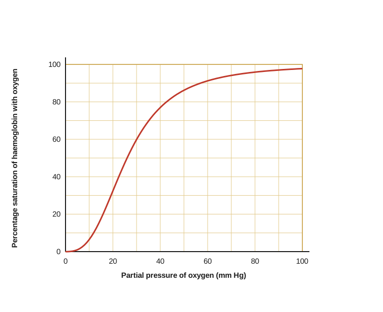
Drawn by us to help you understand the concept clearly, and verified to make sure it's accurate. For exams, practice from your textbook's own diagram.
This figure is the oxygen dissociation curve, and the first thing to notice is its shape: instead of a straight line it sweeps upward as a stretched S, or sigmoid. Read the horizontal direction as the amount of oxygen available in a location and the vertical direction as how fully the haemoglobin is loaded with oxygen. Where oxygen is plentiful, such as in the lungs, the curve is high and flat, so haemoglobin becomes almost completely saturated and picks up its load; where oxygen is scarce, such as in the tissues, the curve drops steeply, so haemoglobin readily lets its oxygen go. Because the binding is reversible, the same pigment grabs oxygen in one place and releases it in another. The curve also lets a student trace how shifting carbon dioxide pressure, risin …