Q.Do you know for how many months pregnancy last in dogs, elephants, cats? Find out.
🔒You're viewing a preview — the full solution, concept, methods & PYQ mapping are locked.
🔒 Start your 14-day free trial to unlock the full solution →Concept understanding — Human Reproduction
Human Reproduction: A First Look
Think about what makes you you. You have your mother's eyes, your father's laugh, a mix of traits from two people you may never have met — your grandparents. That chain of life, stretching back thousands of years, runs through one process: human reproduction. It is not a topic for biology students alone. It is the story of how every single person on this planet came to be here.
The Big Picture: Why Does Reproduction Exist?
Every living thing has one fundamental drive: to continue its kind. A mango tree grows fruit so its seeds can sprout elsewhere. A bacterium splits in two. For humans, reproduction is more complex — and more personal. It is the reason families exist, why you have siblings or cousins, and why populations grow or shrink.
At its simplest, human reproduction is the biological process by which a new human individual is created. It requires two parents — a male and a female — each contributing a special cell that fuses to form a single, new cell. That single cell then grows, divides, and develops into a baby over about nine months.
Reproduction is not the same as sex. Sex is an act; reproduction is the entire sequence from cell fusion to birth. Not every act of sex leads to reproduction, and reproduction can sometimes happen with medical help (like IVF) without sex.
The Two Key Players: Male and Female Reproductive Systems
Your body, whether male or female, has a set of organs designed specifically for reproduction. These are not the same as the organs that keep you alive — they are the "spare set" dedicated to creating the next generation.
The Male System: Production and Delivery
The male reproductive system has one main job: to produce male sex cells (sperm) and deliver them to the female body. The key parts are:
- Testes (two oval organs in a pouch called the scrotum): These are the factories. They produce sperm continuously from puberty onwards — millions every day. They also produce testosterone, the hormone that drives male physical changes at puberty.
- Duct system (a series of tubes): Sperm travel from the testes through a long coiled tube (epididymis) where they mature, then through a muscular tube (vas deferens) that carries them toward the urethra.
- Accessory glands (seminal vesicles, prostate gland, bulbourethral glands): These add fluids to the sperm to nourish them and help them swim. The mixture of sperm and these fluids is called semen.
- Penis: The organ that delivers semen into the female reproductive tract.
The Female System: Production, Reception, and Nurturing
The female system has a bigger job: it produces female sex cells (eggs), receives sperm, and if fertilization occurs, houses and nourishes the developing baby for nine months. The key parts are:
- Ovaries (two almond-sized organs): These produce eggs — but unlike sperm, eggs are not made continuously. A female is born with all the eggs she will ever have (about 1–2 million at birth, reducing to around 300,000–400,000 by puberty). Each month, one egg matures and is released.
- Fallopian tubes (also called oviducts): These are not connected directly to the ovaries. When an egg is released, the finger-like ends of the tube sweep it inside. Fertilization — the meeting of sperm and egg — happens here, in the tube.
- Uterus (womb): A hollow, pear-shaped muscular organ. If fertilization happens, the embryo implants in the lining of the uterus and grows here. If not, the lining is shed — that is menstruation.
- Cervix: The lower, narrow part of the uterus that opens into the vagina. It produces mucus that changes consistency during the month to help or hinder sperm.
- Vagina: A muscular canal that receives the penis during intercourse and serves as the birth canal during delivery.
The female reproductive system has a monthly cycle (the menstrual cycle) that prepares the body for a possible pregnancy. This cycle is controlled by hormones and typically lasts about 28 days. The release of the egg (ovulation) happens around day 14. If no pregnancy occurs, the uterine lining is shed as menstrual blood — this is a normal, healthy process, not a "curse" or something to be ashamed of.
The Moment of Creation: Fertilization
When sperm from the male meet the egg from the female inside a fallopian tube, one sperm may penetrate the egg's outer layer. That single moment — fertilization — creates a new cell called a zygote. This zygote contains 23 chromosomes from the mother and 23 from the father, making 46 in total. That is the complete genetic blueprint for a new human being.
The zygote immediately begins to divide as it travels down the fallopian tube toward the uterus. By the time it reaches the uterus (about 5–7 days later), it has become a ball of about 100 cells called a blastocyst. This blastocyst burrows into the soft, thick lining of the uterus — a process called implantation. That is when pregnancy truly begins.
The Nine-Month Journey: Development
Once implanted, the developing human is called an embryo for the first eight weeks, and then a fetus from the ninth week until birth. During these months:
- A special organ called the placenta develops, connecting the mother's blood supply to the fetus. It delivers oxygen and nutrients and removes waste — without the mother's and baby's blood ever mixing directly.
- The umbilical cord links the baby to the placenta.
- The baby is protected by a fluid-filled sac (amniotic sac) that cushions it.
- All major organs form in the first three months (first trimester). The next three months (second trimester) are about growth and refinement. The final three months (third trimester) are about rapid weight gain and preparation for life outside the womb. …
The NCERT textbook does not answer this itself — it is a "find out" prompt for outside research, not content the exam tests. For reference, typical gestation periods are: dog — about 2 months (~63 days); cat — about 2 months (~63-65 days); elephant — about 18-22 months (~620-660 days), the longest gestation period of any land mammal. …
This is an open "find out for yourself" prompt — NCERT does not state the answer. Real-world gestation periods: dog and cat are both around 2 months, while the elephant's gestation (~18-22 months) is the longest of any land mammal.
This question is explicitly framed by the textbook as something for the student to research independently ("Find out"), immediately after noting that human pregnancy lasts about 9 months — it is meant to prompt a comparison, not to test a memorised NCERT fact, so nothing here is graded exam content.
For reference, the approximate real gestation periods (general zoological knowledge, outside the NCERT text) are:
- Dog: about 58-68 days (roughly 2 months).
- Cat: about 63-65 days (roughly 2 months) — similar to the dog. …
- GUJCET 2026Set 051 markMCQQ.In the given diagram, choose the correct option for labelled 'X' 'Y' and 'Z'. [FIGURE] (A) X - Zona pellucida, Y - Corona radiata, Z - Perivitelline Space (B) X - Corona radiata, Y - Zona pellucida, Z - Perivitelline space (C) X - Zona Pellucida, Y - Perivitelline space, Z - Corona radiata (D) X - Corona radiata, Y - Perivitelline space, Z - Zona pellucida
›Reveal solutionSolution
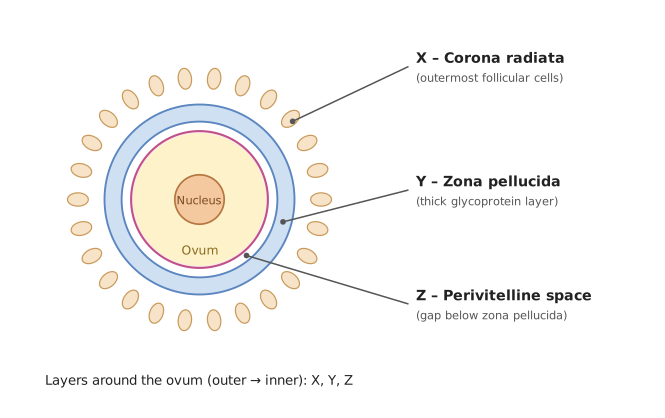
Layers around the ovum from outside inward: X = corona radiata, Y = zona pellucida, Z = perivitelline space (option B). [!TLDR]
Reading the egg's layers from outside to inside gives X = corona radiata, Y = zona pellucida, Z = perivitelline space, i.e. option (B).
Concept
In the secondary oocyte / ovum (NCERT 'Human Reproduction', followed by the GSEB syllabus), the layers surrounding the egg, from the outermost inward, are:
- Corona radiata – the outermost layer of follicular (granulosa) cells.
- Zona pellucida – the thick, transparent glycoprotein membrane beneath the corona radiata. …
- GUJCET 2024Set 101 markMCQQ.What 'X' shows in the given diagram? [FIGURE: female reproductive system diagram with label X pointing to the funnel-shaped end of the oviduct near the ovary] (A) Ampulla (B) Isthmus (C) Infundibulum (D) Fimbriae
›Reveal solutionSolution
[!TLDR]
The funnel-shaped proximal end of the oviduct lying close to the ovary is the infundibulum.
Concept
The human oviduct (fallopian tube) has three regions from the ovarian end towards the uterus: the funnel-shaped infundibulum, the wide ampulla, and the narrow isthmus. The infundibulum bears finger-like fimbriae at its margin that help collect the released ovum.
Solution
The label X points to the funnel-shaped mouth of the oviduct situated near the ovary.
- The fimbriae are only the finger-like fringes at the rim, not the funnel itself. …
- GUJCET 2023Set 071 markMCQQ.During Human fertilisation, this induces changes and acts as a barrier and ensure that only one sperm can enter and fertilise the ovum. (A) Perivitelline space (B) Zona pellucida (C) Corona radiata (D) Cytoplasm of the ovum
›Reveal solutionSolution
On sperm entry, the zona pellucida hardens (zona reaction) and acts as the barrier preventing additional sperm — the block to polyspermy.
Concept — fertilisation and polyspermy block. The secretions on sperm contact induce changes in the zona pellucida, blocking the entry of other sperm so that only one sperm fertilises the …
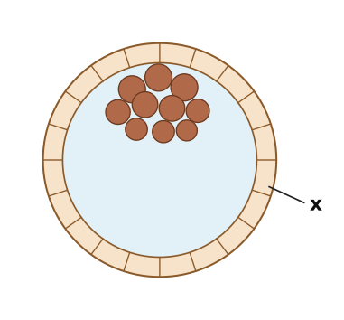
- GUJCET 2022Set 171 markMCQQ.What indicates 'x' in the following diagram? [FIGURE] (A) Trophoblast (B) Inner cell mass (C) Blastomeres (D) Uterine cells
›Reveal solutionSolution
'x' points to the outer single-cell layer of the blastocyst = the trophoblast.
The diagram is a blastocyst. Its outer single layer of cells is the trophoblast (which later aids implantation); the dense inner cluster of cells at one pole is the inner cell mass, and the space is the blastocoel. Since 'x' is on the peripheral wall layer, it is the trophoblast. …
- GUJCET 2021Set 151 markMCQQ.Select the correct option for seasonal breeders. (A) monkeys, dogs (B) dogs, sheep (C) human, tiger (D) human, apes
›Reveal solutionSolution
Seasonal breeders reproduce only in specific favourable seasons.
Concept: Animals are grouped as continuous breeders (reproduce throughout the year) or seasonal breeders (only in particular seasons). Humans, apes, monkeys and tiger are essentially continuous/ye …
- GUJCET 2021Set 151 markMCQQ.The major features of embryonic development at various months of pregnancy are given below. Choose the correct option for correct sequential events. I) The first movements of the foetus. II) The foetus develops limbs and digits. III) The embryo's heart is formed. IV) The body is covered with fine hair. (A) (II), (IV), (I), (III) (B) (IV), (II), (I), (III) (C) (II), (III), (IV), (I) (D) (III), (II), (I), (IV)
›Reveal solutionSolution
[!TLDR] Correct sequence = (III), (II), (I), (IV) → (D).
Concept
Human gestation of about nine months has well-marked developmental milestones. (NCERT Human Reproduction.)
Solution
- After ~1 month: the embryo's heart is formed → III (first).
- By the end of the 2nd month: the foetus develops limbs and digits → II. …
- GUJCET 2020Set x1 markMCQQ.Foetus develop its limbs and digits in..............during embryonic development. (A) Four weeks (B) Eight weeks (C) Twelve weeks (D) Two weeks
›Reveal solutionSolution
Limbs and digits develop by the end of the second month (8 weeks).
During human embryonic development (NCERT, Human Reproduction):
- End of 1 month: heart is formed.
- End of 2 months (8 weeks): the foetus develops limbs and digits. …
🎓Unlock everything free for 14 days
- ✓Full step-by-step solutions
- ✓Concept-first explanations
- ✓Methods, shortcuts & mistakes
- ✓PYQ mapping + timed mock tests
Full access for 14 days. No credit card required.