Q.Draw a labelled diagram of T.S. of Dicot stem. OR
You're viewing a preview — the full solution, concept, methods & PYQ mapping are locked.
Start your 14-day free trial to unlock the full solution →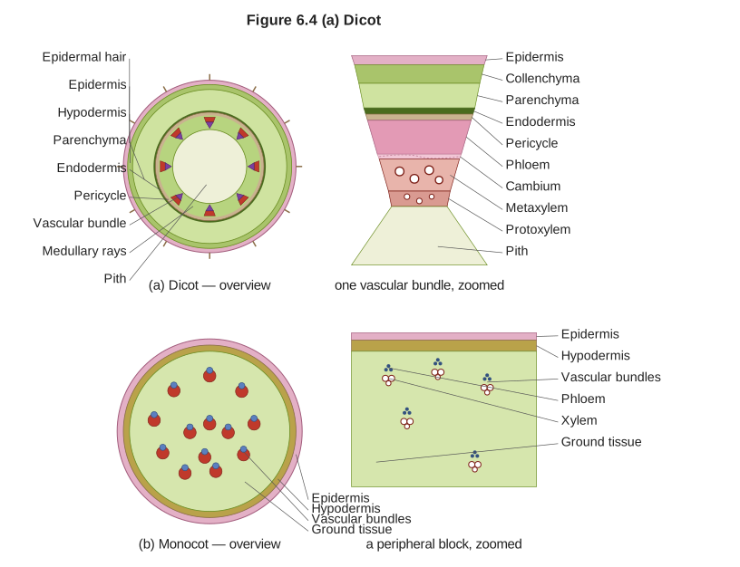
A dicot stem in T.S. shows, from outside to inside: epidermis, hypodermis, cortex, endodermis, pericycle, a ring of open conjoint vascular bundles, and a central pith.
A text-only description is given here (a hand-drawn figure cannot be produced in this format), describing every layer a labelled T.S. of dicot stem diagram should show, from the outermost to the innermost:
-
Epidermis: The outermost single layer of cells, covered by a waxy cuticle; may bear multicellular hairs (trichomes) and stomata.
-
Hypodermis: A few layers just beneath the epidermis, typically made of collenchyma cells, providing mechanical support while allowing some flexibility (unlike the sclerenchymatous hypodermis of monocot stems).
-
General cortex: Parenchymatous cells beneath the hypodermis, often with intercellular spaces; may contain chloroplasts in the outer layers.
-
Endodermis: The innermost layer of the cortex, often called the 'starch sheath' in dicot stems because its cells are typically rich in starch grains.
-
Pericycle: A layer of cells just inside the endodermis, often sclerenchymatous, forming a discontinuous or continuous ring around the vascular bundles.
-
Vascular bundles: Arranged in a distinct ring, each bundle is conjoint (xylem and phloem together on the same radius), collateral (phloem only on the outer side of xylem), and open (a strip of cambium lies between the xylem and phloem, enabling secondary growth). Within each bundle, protoxylem lies towards the centre (pith) and metaxylem towards the periphery — i.e., xylem is endarch.
…
Unlock everything free for 14 days
- Full step-by-step solutions
- Concept-first explanations
- Methods, shortcuts & mistakes
- PYQ mapping + timed mock tests
Full access for 14 days. No credit card required.