Question of 53
Q.Draw a well labelled diagram of the animal cell as seen under electron microscope.
Jammu Kashmir JkboseJammu and Kashmir Board of School Education (Class 11) 2021Subjective· 5mImportance★★★★★
0% · 0/53 Questions
You're viewing a preview — the full solution, concept, methods & PYQ mapping are locked.
Start your 14-day free trial to unlock the full solution →Draw a rounded cell bounded by the plasma membrane, filled with cytoplasm, containing a nucleus and membrane-bound organelles — ER, ribosomes, Golgi, mitochondria, lysosomes, centrioles, etc.; label each part. An animal cell has no cell wall, plastids or large vacuole.
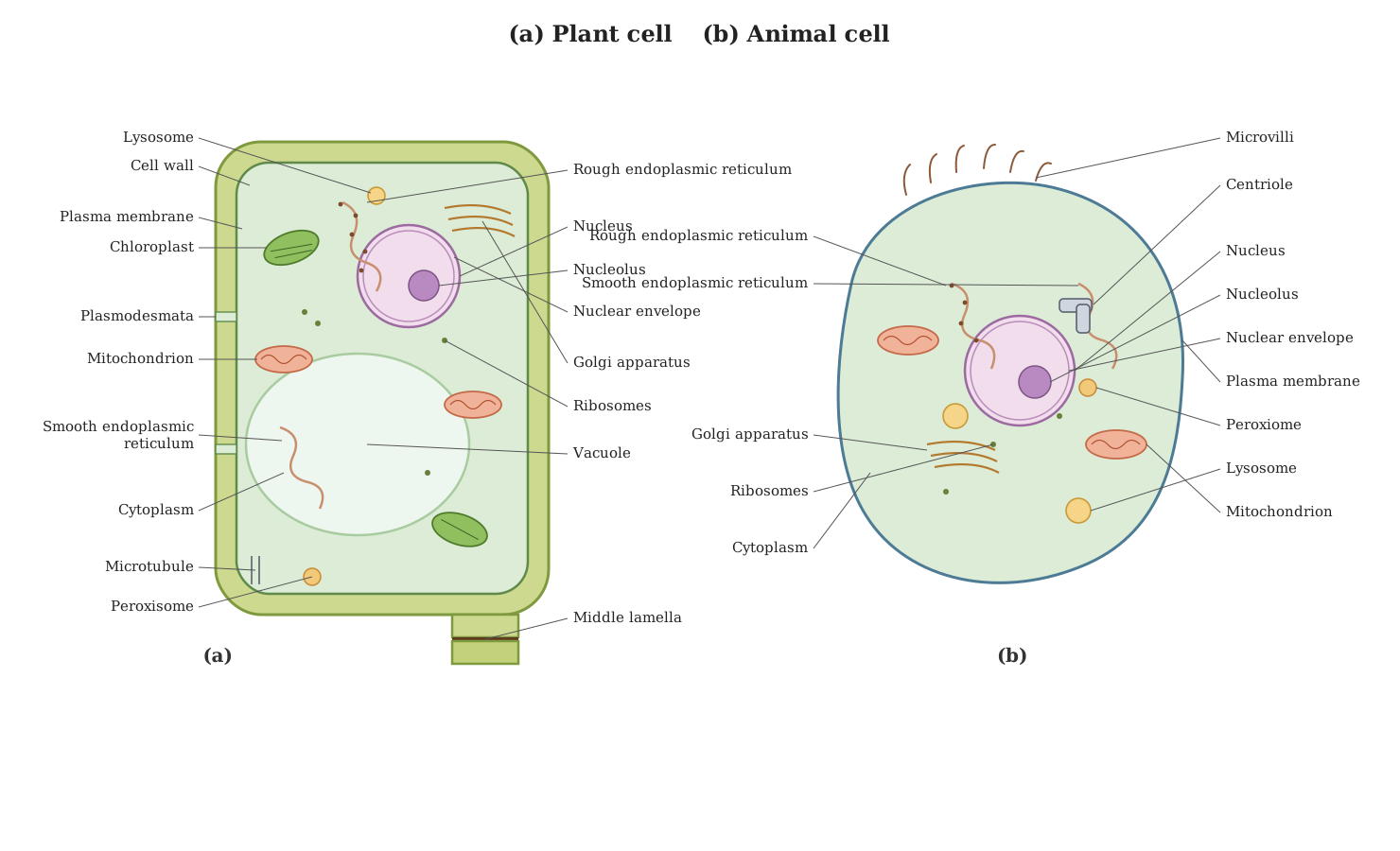
Under the electron microscope the fine (ultra)structure of an animal cell can be seen. A well-labelled diagram should include and clearly label the following parts:
Boundary and ground substance:
- Plasma membrane (cell membrane): the outer living limiting membrane; may show microvilli as finger-like folds.
- Cytoplasm: the ground substance in which organelles are suspended.
Nucleus (label its parts):
- Nuclear envelope with nuclear pores
- Nucleoplasm, chromatin (chromatin network) and nucleolus.
Cytoplasmic organelles to label:
- Endoplasmic reticulum — both rough ER (with ribosomes) and smooth ER.
- Ribosomes (free and on RER).
- Golgi apparatus (Golgi complex).
- Mitochondria (show the inner folds — cristae).
- Lysosomes.
- Centrosome with a pair of centrioles.
- Peroxisomes / microbodies.
- Cytoskeleton (microtubules and microfilaments). …
Unlock everything free for 14 days
- Full step-by-step solutions
- Concept-first explanations
- Methods, shortcuts & mistakes
- PYQ mapping + timed mock tests
Full access for 14 days. No credit card required.