Biology · Ch 18 — Neural Control and Coordination
Neuron as Structural and Functional Unit of Neural System
Neuron as Structural and Functional Unit of Neural System
The neuron is the structural and functional unit of the neural system — a microscopic cell specialised to detect, carry and transmit impulses. A neuron is built from three major parts: the cell body, the dendrites and the axon.
Parts of a neuron
- Cell body (cyton): contains the cytoplasm with the usual cell organelles, along with characteristic granular structures called Nissl's granules.
- Dendrites: short fibres that branch repeatedly and project out from the cell body. They also contain Nissl's granules, and they carry impulses towards the cell body.
- Axon: a single long fibre whose far (distal) end is branched. Each branch ends in a bulb-like swelling called a synaptic knob. The synaptic knobs contain synaptic vesicles, which hold chemicals called neurotransmitters. The axon carries nerve impulses away from the cell body towards a synapse (the junction with the next neuron) or towards a neuro-muscular junction (the junction with a muscle).
Classification by number of axon and dendrites
Based on how many axons and dendrites a neuron possesses, neurons are grouped into three types:
- Multipolar — one axon together with two or more dendrites; found, for example, in the cerebral cortex.
- Bipolar — one axon and a single dendrite; found, for example, in the retina of the eye.
- Unipolar — the cell body carries only one axon (no true dendrites); this type is usually seen in the embryonic stage.
Two types of axons
Axons are of two kinds, depending on whether they are wrapped in a myelin sheath: …
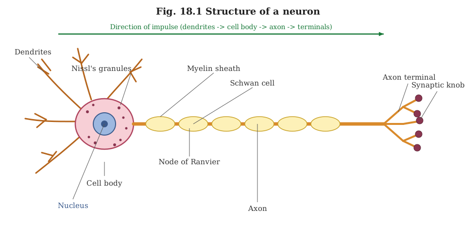
Drawn by us to help you understand the concept clearly, and verified to make sure it's accurate. For exams, practice from your textbook's own diagram.
This diagram shows the three regions that make up a typical neuron and how they fit together into one conducting cell. At the centre is the cell body, or cyton — a rounded mass of cytoplasm holding the nucleus and the granular Nissl's bodies; it is the metabolic hub of the neuron. Radiating from it are several short, branching dendrites, which receive signals and carry impulses inward toward the cyton. From the opposite side arises a single long process, the axon, which carries the impulse away from the cell body. Along the axon the diagram marks the myelin sheath, an insulating covering laid down in segments, with small unwrapped gaps between segments called the nodes of Ranvier; this segmented insulation lets the impulse jump from node to node and travel faster. The axon finally divides into fine branches, each tipped with a swollen synaptic knob (axon terminal) through which th …