Q.Do you know for how many months pregnancy last in dogs, elephants, cats? Find out.
🔒You're viewing a preview — the full solution, concept, methods & PYQ mapping are locked.
🔒 Start your 14-day free trial to unlock the full solution →Concept understanding — Human Reproduction
Human Reproduction: A First Look
Think about what makes you you. You have your mother's eyes, your father's laugh, a mix of traits from two people you may never have met — your grandparents. That chain of life, stretching back thousands of years, runs through one process: human reproduction. It is not a topic for biology students alone. It is the story of how every single person on this planet came to be here.
The Big Picture: Why Does Reproduction Exist?
Every living thing has one fundamental drive: to continue its kind. A mango tree grows fruit so its seeds can sprout elsewhere. A bacterium splits in two. For humans, reproduction is more complex — and more personal. It is the reason families exist, why you have siblings or cousins, and why populations grow or shrink.
At its simplest, human reproduction is the biological process by which a new human individual is created. It requires two parents — a male and a female — each contributing a special cell that fuses to form a single, new cell. That single cell then grows, divides, and develops into a baby over about nine months.
Reproduction is not the same as sex. Sex is an act; reproduction is the entire sequence from cell fusion to birth. Not every act of sex leads to reproduction, and reproduction can sometimes happen with medical help (like IVF) without sex.
The Two Key Players: Male and Female Reproductive Systems
Your body, whether male or female, has a set of organs designed specifically for reproduction. These are not the same as the organs that keep you alive — they are the "spare set" dedicated to creating the next generation.
The Male System: Production and Delivery
The male reproductive system has one main job: to produce male sex cells (sperm) and deliver them to the female body. The key parts are:
- Testes (two oval organs in a pouch called the scrotum): These are the factories. They produce sperm continuously from puberty onwards — millions every day. They also produce testosterone, the hormone that drives male physical changes at puberty.
- Duct system (a series of tubes): Sperm travel from the testes through a long coiled tube (epididymis) where they mature, then through a muscular tube (vas deferens) that carries them toward the urethra.
- Accessory glands (seminal vesicles, prostate gland, bulbourethral glands): These add fluids to the sperm to nourish them and help them swim. The mixture of sperm and these fluids is called semen.
- Penis: The organ that delivers semen into the female reproductive tract.
The Female System: Production, Reception, and Nurturing
The female system has a bigger job: it produces female sex cells (eggs), receives sperm, and if fertilization occurs, houses and nourishes the developing baby for nine months. The key parts are:
- Ovaries (two almond-sized organs): These produce eggs — but unlike sperm, eggs are not made continuously. A female is born with all the eggs she will ever have (about 1–2 million at birth, reducing to around 300,000–400,000 by puberty). Each month, one egg matures and is released.
- Fallopian tubes (also called oviducts): These are not connected directly to the ovaries. When an egg is released, the finger-like ends of the tube sweep it inside. Fertilization — the meeting of sperm and egg — happens here, in the tube.
- Uterus (womb): A hollow, pear-shaped muscular organ. If fertilization happens, the embryo implants in the lining of the uterus and grows here. If not, the lining is shed — that is menstruation.
- Cervix: The lower, narrow part of the uterus that opens into the vagina. It produces mucus that changes consistency during the month to help or hinder sperm.
- Vagina: A muscular canal that receives the penis during intercourse and serves as the birth canal during delivery.
The female reproductive system has a monthly cycle (the menstrual cycle) that prepares the body for a possible pregnancy. This cycle is controlled by hormones and typically lasts about 28 days. The release of the egg (ovulation) happens around day 14. If no pregnancy occurs, the uterine lining is shed as menstrual blood — this is a normal, healthy process, not a "curse" or something to be ashamed of.
The Moment of Creation: Fertilization
When sperm from the male meet the egg from the female inside a fallopian tube, one sperm may penetrate the egg's outer layer. That single moment — fertilization — creates a new cell called a zygote. This zygote contains 23 chromosomes from the mother and 23 from the father, making 46 in total. That is the complete genetic blueprint for a new human being.
The zygote immediately begins to divide as it travels down the fallopian tube toward the uterus. By the time it reaches the uterus (about 5–7 days later), it has become a ball of about 100 cells called a blastocyst. This blastocyst burrows into the soft, thick lining of the uterus — a process called implantation. That is when pregnancy truly begins.
The Nine-Month Journey: Development
Once implanted, the developing human is called an embryo for the first eight weeks, and then a fetus from the ninth week until birth. During these months:
- A special organ called the placenta develops, connecting the mother's blood supply to the fetus. It delivers oxygen and nutrients and removes waste — without the mother's and baby's blood ever mixing directly.
- The umbilical cord links the baby to the placenta.
- The baby is protected by a fluid-filled sac (amniotic sac) that cushions it.
- All major organs form in the first three months (first trimester). The next three months (second trimester) are about growth and refinement. The final three months (third trimester) are about rapid weight gain and preparation for life outside the womb. …
The NCERT textbook does not answer this itself — it is a "find out" prompt for outside research, not content the exam tests. For reference, typical gestation periods are: dog — about 2 months (~63 days); cat — about 2 months (~63-65 days); elephant — about 18-22 months (~620-660 days), the longest gestation period of any land mammal. …
This is an open "find out for yourself" prompt — NCERT does not state the answer. Real-world gestation periods: dog and cat are both around 2 months, while the elephant's gestation (~18-22 months) is the longest of any land mammal.
This question is explicitly framed by the textbook as something for the student to research independently ("Find out"), immediately after noting that human pregnancy lasts about 9 months — it is meant to prompt a comparison, not to test a memorised NCERT fact, so nothing here is graded exam content.
For reference, the approximate real gestation periods (general zoological knowledge, outside the NCERT text) are:
- Dog: about 58-68 days (roughly 2 months).
- Cat: about 63-65 days (roughly 2 months) — similar to the dog. …
- KCET 2025Set C-41 markMCQQ.Select the mismatched pair :(1) First month of pregnancy – Formation of heart(2) Second month of pregnancy – Movement of foetus(3) Third month of pregnancy – Formation of most of the major organ systems(4) Six month of pregnancy – Eye lids separate and eye lashes are found (A) b (B) c (C) d (D) a
›Reveal solutionSolution
Check each month against the NCERT embryonic time-line; only the 'second month → movement of foetus' pairing is wrong, because the first foetal movements appear in the fifth month.
The four listed pairs correspond to a, b, c, d respectively.
-
(a) First month – Formation of heart. ✓ Correct. "After one month of pregnancy, the embryo's heart is formed."
-
(b) Second month – Movement of foetus. ✗ Mismatched. By the end of the second month the foetus develops limbs and digits. The first movements of the foetus (together with the appearance of hair on the head) are observed during the fifth month. So pairing month 2 with foetal movement is incorrect. …
-
- KCET 2025Set C-41 markMCQQ.In which of the following, HIV replicates and produces its progeny viruses? (A) Memory T-lymphocytes (B) Killer T-lymphocytes (C) Suppressor T-lymphocytes (D) Helper T-lymphocytes
›Reveal solutionSolution
HIV targets the CD4-bearing helper T cell, which becomes the virus factory — hence the progressive fall in helper T counts in AIDS.
Step 1 — The route of infection.
After entering the body, HIV first enters macrophages, where its RNA genome is copied into DNA by reverse transcriptase; this viral DNA integrates into the host cell's DNA and directs the production of virus particles. The macrophage keeps functioning as an HIV factory.
Step 2 — The key target cell.
HIV then enters helper T-lymphocytes (TH cells) — it does so because its envelope glycoprotein binds the CD4 receptor, and helper T cells are the CD4⁺ population. Inside them, the virus replicates and produces progeny viruses, which are released into the blood and go on to attack further helper T cells.
Step 3 — Why this matters clinically (the internal check). …
- KCET 2024Set B-41 markMCQQ.In human females, the endometrium of uterus consists of (A) Smooth muscle (B) Glandular layer (C) Adipose layer (D) Cartilaginous layer
›Reveal solutionSolution
Endometrium = the inner glandular lining of the uterus (the layer that cycles and is shed in menstruation); the muscle layer is the myometrium.
Step 1 — The three layers of the uterine wall
Layer Position Tissue Perimetrium Outer Thin serous/membranous covering Myometrium Middle Thick smooth muscle — its strong contractions drive parturition Endometrium Inner Glandular mucosal epithelium lining the uterine cavity Step 2 — Why the endometrium is glandular
The endometrium is the functional, hormone-responsive lining:
- Under estrogen it proliferates (proliferative phase).
- Under progesterone from the corpus luteum its glands become secretory, storing glycogen to nourish an arriving blastocyst (secretory phase).
- It is the layer in which implantation occurs.
- If fertilisation fails, the corpus luteum degenerates, progesterone falls, and the endometrium breaks down and is shed — this is menstruation. …
- KCET 2023Set B-41 markMCQQ.With reference to human sperm, match the List-I with List-II. List-I | List-II
- Head | p. Filled with enzyme
- Acrosome | q. Contains mitochondria
- Middle piece | r. Sperm motility
- Tail | s. Contains haploid nucleus
›Reveal solutionSolution
Recall sperm structure: head = haploid nucleus, acrosome = enzyme-filled cap, middle piece = mitochondria, tail = motility.
Concept — Structure of the human spermatozoon (Human Reproduction). A sperm is a microscopic haploid cell made of a head, neck, middle piece and tail, all covered by a plasma membrane.
-
Head — contains an elongated haploid nucleus carrying the paternal genome. → s
-
Acrosome — a cap-like structure over the anterior part of the head, formed from the Golgi body and filled with hydrolytic enzymes (e.g. hyaluronidase) that help the sperm penetrate the zona pellucida of the ovum at fertilisation. → p
-
Middle piece — possesses numerous mitochondria arranged in a spiral sheath; these generate the ATP that powers the flagellar beat. → q …
- KCET 2022Set A-11 markMCQQ.In which month of gestation, the first movements of foetus and appearance of hair on its head is observed ? (A) 5th month (B) 1st month (C) 8th month (D) 4th month
›Reveal solutionSolution
The first foetal movements (quickening) and the appearance of hair on the head both typically occur around the 5th month of gestation, making option (A) the correct choice.
The question asks about two specific developmental milestones: when the mother first feels the foetus move, and when hair begins to appear on the foetus’s head. These are standard landmarks in prenatal development, and they are often tested together because they occur at roughly the same time.
In human gestation, the first movements of the foetus — called "quickening" — are usually felt by the mother between the 4th and 5th month. However, the first clearly noticeable movements are most consistently reported around the 5th month (18–20 weeks). At this stage, the foetus is large enough and its muscles strong enough for the movements to be perceptible.
Simultaneously, the development of hair on the head (lanugo, which later becomes finer hair, and then true scalp hair) begins around the same period. By the end of the 5th month, fine hair is visible on the head, and this is the standard textbook milestone.
Let’s walk through the reasoning step by step.
-
First foetal movements
The foetus starts moving much earlier (around 8–10 weeks), but these movements are too weak for the mother to feel. By the 5th month (18–20 weeks), the foetus has grown enough that its limb movements become strong and are felt by the mother. This is a classic milestone in obstetrics.
-
Appearance of hair on the head
Hair follicles begin forming around the 3rd month, but visible hair on the scalp appears later. By the 5th month, the foetus has a fine layer of hair (lanugo) covering the body, and hair on the head becomes distinct. This is the accepted timing in standard biology curricula.
-
Checking the options
- (A) 5th month: Matches both milestones. …
-
- KCET 2022Set A-11 markMCQQ.Maintenance of constant internal environment is called as (A) Thermoregulation (B) Metastasis (C) Osmoregulation (D) Homeostasis
›Reveal solutionSolution
"Maintenance of a constant internal environment" is the textbook definition of homeostasis; the other options are either sub-processes of it or unrelated.
Step 1 — The definition. The NCERT chapter Organisms and Populations opens with exactly this idea: organisms that cannot tolerate a changing internal state regulate and thereby maintain homeostasis — constancy of the internal environment — even when the external conditions (temperature, salinity, pH, water availability) fluctuate.
Step 2 — Examples. Mammals and birds keep their body temperature near 37∘C by sweating/shivering; the kidney holds blood osmolarity steady; insulin and glucagon hold blood glucose steady. Each is a homeostatic mechanism, and each works by negative feedback.
Step 3 — Why the other options are narrower or wrong. …
- KCET 2021Set C-31 markMCQQ.In the given diagram of human sperm, identify the functions of the labelled parts. a, b and c. 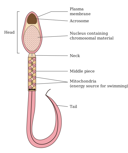 (A) a-Helps in penetration of sperm into ovum, b-Helps in movement of sperm, c-Provides energy for the movement of sperms into the female reproductive tract. (B) a-Helps in penetration of sperm into ovum, b-Provides energy for the movement of sperm, c-Helps in movement of sperm. (C) a-Helps in movement of sperm, b-Helps in penetration of sperm into ovum, c-Provides energy for the movement of sperms (D) a-Provides energy for the movement of sperm, b-Helps in movement of sperm, c-Helps in penetration of sperm into ovum
›Reveal solutionSolution
Head = penetration (acrosome), middle piece = energy (mitochondria), tail = movement (flagellum) — the standard three-region sperm structure.
Region a — the head.
Contains the acrosome, a cap of enzymes that digests the egg's outer layers — its function is to help the sperm penetrate the ovum.
Region b — the middle piece.
Densely packed with mitochondria, spiralled around the axial filament — its function is to provide the energy (ATP) the tail needs to keep moving. …
- KCET 2020Set A-11 markMCQQ.Match the months listed in Column-I with the organogenesis of foetus in Column-II. Column-I Column-II(i) First month(a) Separation of eye lids(ii) Second month(b) Hairs on head(iii) Fifth month(c) Heart(iv) Six month(d) Limbs & digits (A)(c)(d)(a)(b) (B)(b)(c)(d)(a) (C)(d)(b)(c)(a) (D)(c)(d)(b) (a)
›Reveal solutionSolution
This question tests the timeline of human foetal development. The correct matching is: First month → Heart, Second month → Limbs & digits, Fifth month → Hairs on head, Sixth month → Separation of eyelids. That corresponds to option (D).
The key here is to recall the sequence of organogenesis — the process by which organs form and mature in the developing foetus. Each month brings a new set of milestones, and the order is logical: the most vital organs (like the heart) form first, followed by the body's structure (limbs, digits), then surface features (hair), and finally fine details (eyelid separation).
Let’s match each month to its landmark event.
-
First month (i) → (c) Heart
By the end of the first month (week 4), the heart is the first functional organ to form and begins beating. This is the earliest major organogenesis event among the options.
-
Second month (ii) → (d) Limbs & digits
During the second month (weeks 5–8), the limb buds appear and differentiate into arms, legs, fingers, and toes. This is when the basic body plan takes shape.
-
Fifth month (iii) → (b) Hairs on head
By the fifth month (around week 20), lanugo (fine hair) covers the body, and scalp hair begins to grow. This is a later developmental feature. …
-
- KCET 2020Set A-11 markMCQQ.In the life cycle of plasmodium, fertilisation takes place in (A) Salivary glands of mosquito (B) RBCs of humans (C) Stomach of mosquito (D) Liver cells
›Reveal solutionSolution
The key is that Plasmodium completes its sexual cycle (including fertilisation) inside the mosquito’s gut, not in the human host. The correct site is the stomach of the mosquito.
The life cycle of Plasmodium (the malarial parasite) alternates between a human host and a female Anopheles mosquito. The critical distinction to remember is that sexual reproduction (gametogony and fertilisation) happens only inside the mosquito, while asexual reproduction (schizogony) happens inside the human. Many students mistakenly think fertilisation occurs in human blood because they see gametocytes there — but those gametocytes are merely the precursors; they must be taken up by the mosquito to complete the sexual phase.
Let’s trace the sequence:
-
In humans (asexual cycle)
When an infected mosquito bites a human, it injects sporozoites into the blood. These travel to the liver, invade liver cells, multiply, and then burst out as merozoites. The merozoites invade red blood cells (RBCs), where they multiply further, causing the cyclical fevers. Some merozoites, instead of continuing asexual multiplication, develop into gametocytes (male and female) inside RBCs. These gametocytes circulate in the blood but do not fuse inside the human — they are dormant until taken up by a mosquito.
-
In the mosquito (sexual cycle)
When a female Anopheles mosquito bites an infected human, it sucks up blood containing these gametocytes. The gametocytes enter the mosquito’s stomach (also called the midgut). Here, the change in environment (lower temperature, higher pH) triggers the gametocytes to mature into gametes. The male gamete (microgamete) fertilises the female gamete (macrogamete) inside the stomach lumen — this is the fertilisation event.
-
After fertilisation …
-
- KCET 2018Set A-11 markMCQQ.In the following reaction, identify X and Y respectively: Pyruvic acid + CoA + NAD+ Mg2+ X → Y + CO2 + NADH + H+ (A) Water, Acetyl CoA (B) Acetyl CoA, Pyruvate dehydrogenase (C) Pyruvate dehydrogenase, Acetyl CoA (D) Pyruvate dehydrogenase, Oxalo-acetic acid
›Reveal solutionSolution
The link (transition) reaction is pyruvate+CoA+NAD+pyruvate dehydrogenaseacetyl CoA+CO2+NADH+H+ — so X is the enzyme complex and Y is acetyl CoA.
Step 1 — Locate the reaction.
After glycolysis, pyruvic acid formed in the cytoplasm enters the mitochondrial matrix. There it undergoes oxidative decarboxylation — the "link reaction" that connects glycolysis to the Krebs cycle.
Step 2 — The complete equation.
Pyruvic acid+CoA+NAD+pyruvate dehydrogenase complex Mg2+, TPP Acetyl CoA+CO2+NADH+H+
The reaction is catalysed by the pyruvate dehydrogenase complex, which requires several cofactors — notably Mg2+ (shown over the arrow in the stem) and TPP.
Step 3 — Assign X and Y.
In the stem, the reaction is written as reactants → X → Y + CO2 + NADH + H+.
- X is the agent that acts on the reactants — the enzyme, pyruvate dehydrogenase.
- Y is the carbon-containing product that goes on to enter the Krebs cycle — acetyl CoA (the 2-carbon acetyl group carried on coenzyme A). …
- KCET 2018Set A-11 markMCQQ.The hormones of "Fright, Fight and Flight" are (A) Thyroxin and Oxytocin (B) Thyroxin and Melatonin (C) Adrenalin and Nor-adrenalin (D) Gastrin and Secretin
›Reveal solutionSolution
The adrenal medulla releases adrenaline and noradrenaline — the emergency hormones that produce the fright/fight/flight response.
Step 1 — Recall the adrenal gland.
Each adrenal gland has two parts: the outer cortex (secretes corticoids — glucocorticoids like cortisol, mineralocorticoids like aldosterone) and the inner medulla (secretes the catecholamines: adrenaline/epinephrine and noradrenaline/norepinephrine).
Step 2 — The emergency response.
Under stress, emergency or excitement, the sympathetic nervous system stimulates the adrenal medulla. The catecholamines released produce a coordinated set of effects that prepare the body to face or escape danger:
- increased heart rate, force of contraction and cardiac output
- increased rate of respiration and dilation of bronchioles
- glycogenolysis and lipolysis, raising blood glucose and free fatty acids to fuel muscles
- pupillary dilation, piloerection, sweating …
🎓Unlock everything free for 14 days
- ✓Full step-by-step solutions
- ✓Concept-first explanations
- ✓Methods, shortcuts & mistakes
- ✓PYQ mapping + timed mock tests
Full access for 14 days. No credit card required.