Q.You can easily study pollen germination by dusting some pollen from flowers such as pea, chickpea, Crotalaria, balsam, and Vinca on a glass slide containing a drop of sugar solution (about 10 per cent). After about 15–30 minutes, observe the slide under the low power lens of the microscope. What do you expect to see?
Concept understanding — Male And Female Gametophyte
Let’s start with something you already know. Think of a plant like a mango tree. It produces flowers, and those flowers eventually turn into mangoes. How does that happen? The flower is the plant’s reproductive organ, and inside it, two tiny but crucial structures are made: the male gametophyte and the female gametophyte. These are not the “male” and “female” parts of the flower themselves (like the stamen and pistil) — they are the microscopic, single-generation cells that actually carry the genetic material to form the next plant.
The everyday intuition
Imagine you are baking a cake. The male gametophyte is like the packet of dry yeast — it’s small, mobile, and needs to be added to the mixture. The female gametophyte is like the bowl of flour, eggs, and sugar — it’s larger, stationary, and provides the environment where the yeast works. Without the yeast reaching the bowl, no cake rises. Without the bowl, the yeast has nothing to act on. In a flower, the male gametophyte (pollen grain) must travel to the female gametophyte (embryo sac) for fertilisation to happen.
The precise meaning
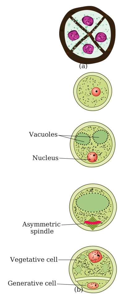
In NCERT biology, the male gametophyte is the pollen grain. It develops inside the anther (the top part of the stamen). A mature pollen grain contains two cells: a tube cell and a generative cell. The tube cell will grow a long tube down the style of the pistil, and the generative cell will divide to form two sperm cells. So the male gametophyte is not the whole stamen — it’s the tiny, single-celled (or two-celled) structure that carries the male genetic material.
The female gametophyte is the embryo sac. It develops inside the ovule, which is located in the ovary of the pistil. The embryo sac is a seven-celled, eight-nucleate structure (though you don’t need to memorise numbers for a prose subject — just know it’s a small, organised sac). It contains the egg cell (the female gamete) and other cells that help in fertilisation and nourishment. The female gametophyte is not the whole pistil — it’s the microscopic sac inside the ovule.
The male gametophyte (pollen grain) is haploid — it has only one set of chromosomes. The female gametophyte (embryo sac) is also haploid. When they fuse during fertilisation, they form a diploid zygote, which grows into the seed. This is why both are called “gametophytes” — they produce gametes (sperm and egg).
Why it matters
Without the male and female gametophytes, no seed would form, and no fruit would develop. In flowering plants, the male gametophyte is the pollen that bees carry, wind blows, or humans collect for pollination. The female gametophyte is the hidden, protected structure inside the ovary. Their meeting is the moment of fertilisation — the start of a new plant’s life.
For a commerce or humanities student, the key takeaway is this: gametophytes are the tiny, single-generation carriers of genetic material in plants. They are not the visible flower parts — they are the microscopic “packages” that ensure reproduction. In NCERT, this concept is the foundation for understanding pollination, fertilisation, and seed formation. It’s a simple but powerful idea: life passes from one generation to the next through these miniature, specialised cells.
Students revising this topic often search for "Male And Female Gametophyte NEET questions", "Male And Female Gametophyte important questions", or "Male And Female Gametophyte class 12 biology". This concept is directly part of the Sexual Reproduction in Flowering Plants chapter in the NCERT/CBSE Class 12 Biology syllabus, and it is also an important topic for NEET and state medical/CET entrance exams, making it worth mastering for both board and competitive-exam preparation.
Under the low-power lens, after 15–30 minutes on the sugar-solution slide, you should see pollen tubes emerging from the pollen grains — each germinated grain puts out a thin tubular outgrowth (through a germ pore) that grows out into the sugar solution, mimicking how a pollen tube would grow down the style toward the ovule after real pollination.
Pollen tubes growing out of the germinated pollen grains.
On a 10% sugar-solution slide, viable pollen grains germinate within 15-30 minutes and put out visible pollen tubes — this practical directly demonstrates the first step of pollen-pistil interaction (§1.2.3) outside the flower.
Why this works: a sugar solution approximates the sucrose-rich medium the stigma surface naturally provides, which stimulates a compatible, viable pollen grain to germinate — the same trigger that starts pollen-tube growth in vivo after pollination.
What you should observe under the low-power lens:
- Most pollen grains will appear as small, roughly spherical bodies on the slide (their genus-specific exine ornamentation may be visible).
- From a proportion of the grains, a thin pollen tube emerges through one of the grain's germ pores and grows outward into the sugar solution.
- The pollen tube carries the grain's two male gametes (the vegetative cell guiding the tube's growth, with the generative cell/its two sperm cells inside it) — exactly as it would when growing through the style toward the micropyle of an ovule in a real pollinated flower.
This is why the activity is a useful, simple classroom proxy for pollen-pistil interaction: it isolates and makes directly visible the germination + tube-growth step that is otherwise hidden inside the pistil.
Pollen tubes growing out of the germinated pollen grains — visible proof that the pollen was viable and has begun the same tube-growth process it would use inside a real pistil.
Showing the 12 most recent of 21 on this concept.
- KCET 2025Set C-41 markMCQQ.When pollen grains of a flower of a plant pollinate the stigma of flower of another plant, it is called (A) Autogamy (B) Dichogamy (C) Geitonogamy (D) Xenogamy
›Reveal solutionSolution
Pollen from one plant reaching the stigma of a flower on another plant is xenogamy — the only type that brings genetically different pollen to the stigma.
Step 1 — The three kinds of pollination
- Autogamy — pollen is transferred from the anther to the stigma of the same flower. Requires anther and stigma to lie close and to mature together (synchrony). Chasmogamous flowers do it partly; cleistogamous flowers (which never open) do it obligatorily.
- Geitonogamy — pollen goes to another flower of the same plant. Functionally it needs a pollinator (so it looks like cross-pollination), but genetically it is still autogamy, because all the flowers of one plant carry the same genes.
- Xenogamy — pollen is carried to the stigma of a flower on a different plant. This is the only type that brings genetically different pollen to the stigma, and hence the only true cross-pollination.
Step 2 — Match the stem
The stem says: pollen of a flower of a plant pollinates the stigma of a flower of another plant. Two different plants ⇒ xenogamy.
Step 3 — Eliminate the trap
Dichogamy (option B) is not a type of pollination at all — it is the device by which a plant prevents self-pollination, i.e. the anther and stigma mature at different times. It is a distractor.
✓Final answerThe correct option is (D) — Xenogamy.
ANSWER: D
- KCET 2025Set C-41 markMCQQ.:
 Which of these options is true in the context of the above diagram of pollen grain ?
(A) 'A' is a vegetative cell which gives rise to male gametes and 'B' is a generative cell which produces pollen tube
(B) 'A' is a generative cell which gives rise to pollen tube and 'B' is a vegetative cell which form male gametes
(C) 'A' is a vegetative cell with abundant food reserve and 'B' is a generative cell which form male gametes
(D) 'A' is a generative cell which forms male gametes and 'B' is a vegetative cell which produces pollen tube
Which of these options is true in the context of the above diagram of pollen grain ?
(A) 'A' is a vegetative cell which gives rise to male gametes and 'B' is a generative cell which produces pollen tube
(B) 'A' is a generative cell which gives rise to pollen tube and 'B' is a vegetative cell which form male gametes
(C) 'A' is a vegetative cell with abundant food reserve and 'B' is a generative cell which form male gametes
(D) 'A' is a generative cell which forms male gametes and 'B' is a vegetative cell which produces pollen tube›Reveal solutionSolution
Identify the two cells by size and shape: the big food-laden cell is the vegetative cell, the small spindle/lens-shaped cell cut off against the wall is the generative cell that makes the male gametes — which is exactly what option (C) states.
Step 1 — Read the diagram.
The drawing is a mature (2-celled) pollen grain: a spiky sculptured outer wall (the exine, made of sporopollenin) with germ pores, and a smooth inner wall (the intine). A single curved line inside divides the cytoplasm into two unequal cells — the state in which most flowering-plant pollen is shed.
- Leader A points to the large region occupying most of the grain.
- Leader B points to the small lens- / crescent-shaped cell lying against the wall.
Step 2 — The biology of the microspore's unequal division.
The microspore divides unequally, giving two very different cells:
Cell Size / shape Contents Function Vegetative (tube) cell Bigger, fills most of the grain Abundant food reserve; large, irregularly shaped nucleus On germination it produces the pollen tube Generative cell Small, spindle- / lens-shaped, floating in the vegetative cell's cytoplasm Dense cytoplasm, little reserve Divides mitotically to form the two male gametes So the two roles are tied to the two sizes: big → vegetative → food store + pollen tube; small → generative → male gametes. (In roughly 60% of species the generative cell divides only after the pollen tube has grown; in the other 40% it has already divided, giving a 3-celled grain at shedding.)
Step 3 — Map onto the labels.
From the figure, A = the large cell = vegetative cell and B = the small lens-shaped cell = generative cell. Hence A carries the abundant food reserve, and B is the one that gives rise to the male gametes.
Step 4 — Test each option.
- (A) Makes the vegetative cell produce the male gametes and the generative cell the pollen tube — the two functions are swapped. ✗
- (B) Calls A generative and B vegetative — sizes swapped and functions swapped. ✗
- (C) A = vegetative cell with abundant food reserve; B = generative cell which forms male gametes — agrees with both the drawing and the biology. ✓
- (D) Calls A generative — but A is the large cell, so this contradicts the figure. ✗
✓Final answerThe correct option is (C) — 'A' is a vegetative cell with abundant food reserve and 'B' is a generative cell which form male gametes.
ANSWER: C
- KCET 2023Set B-41 markMCQQ.A flower has 10 stamens each having bilobed dithecous anther. If each microsporangium has 5 pollen mother cells, how many pollen grains would be produced by the flower? (A) 1600 (B) 200 (C) 400 (D) 800
›Reveal solutionSolution
Count microsporangia (4 per anther), multiply by PMCs per sporangium, then by 4 pollen grains produced per PMC by meiosis.
Step 1 — The anatomy of a typical anther.
A typical angiosperm anther is bilobed (two lobes) and dithecous (each lobe has two theca). It is therefore tetrasporangiate — it contains four microsporangia, two located in each lobe, at the corners in a transverse section.
microsporangia per anther=4
Step 2 — Total microsporangia in the flower.
Each stamen bears one anther, and there are 10 stamens:
10 stamens×4 anthermicrosporangia=40 microsporangia
Step 3 — Total pollen mother cells (microspore mother cells).
The question gives 5 PMCs per microsporangium (a simplifying figure — in reality there are many more):
40×5=200 PMCs
Step 4 — Apply meiosis: the key biology.
Each pollen mother cell is diploid and undergoes meiosis (microsporogenesis). One meiotic division of one cell yields four haploid microspores, arranged as a tetrad. Each microspore matures into one pollen grain.
1 PMC meiosis 4 microspores=4 pollen grains
Step 5 — Total pollen grains.
200 PMCs×4=800 pollen grains
Checking the trap options: 200 is the PMC count (forgetting meiosis); 400 comes from wrongly assuming a bilobed anther has only 2 microsporangia; 1600 comes from double-counting (e.g. treating each of the 10 stamens as bearing 2 anthers). The complete chain is:
10×4×5×4=800
✓Final answerThe correct option is (D) — 800.
ANSWER: D
- KCET 2023Set B-41 markMCQQ.Primary endosperm nucleus is formed by fusion of (A) Two polar nuclei and two male gametes (B) Two polar nuclei and one male gamete (C) Ovum and male gamete (D) One polar nucleus and male gamete
›Reveal solutionSolution
The primary endosperm nucleus arises by triple fusion — one male gamete + the two polar nuclei of the central cell — making it triploid (3n).
1. The setting: what the pollen tube delivers
A mature male gametophyte (pollen grain) delivers two male gametes into the embryo sac. The female gametophyte it enters is the 7-celled, 8-nucleate embryo sac, in which the important players are:
- the egg cell (with 2 synergids, forming the egg apparatus), and
- the large central cell, which contains two polar nuclei.
Because both male gametes are used up in two separate fusion events, the process is called double fertilisation — a feature unique to angiosperms.
2. The two fusions
- Syngamy — one male gamete fuses with the egg cell:
n (male gamete)+n (egg)⟶2n zygote
- Triple fusion — the other male gamete moves to the central cell and fuses with the two polar nuclei:
The name "triple fusion" itself is the memory hook: three nuclei are involved, and the product is triploid. The central cell now becomes the primary endosperm cell (PEC), which divides to form the nutritive endosperm that feeds the developing embryo.
n (male gamete)+n+n (two polar nuclei)⟶3n Primary Endosperm Nucleus (PEN)
3. Why the other options fail
- (A) Two polar nuclei and two male gametes — this would use up both male gametes on the central cell, leaving nothing to fertilise the egg, and would give a tetraploid (4n) nucleus. Wrong.
- (C) Ovum and male gamete — that fusion is syngamy, and its product is the zygote (2n), not the PEN.
- (D) One polar nucleus and male gamete — this would be a double fusion giving 2n; but both polar nuclei participate, and the PEN is 3n.
✓Final answerThe correct option is (B) — Two polar nuclei and one male gamete.
ANSWER: B
- KCET 2023Set B-41 markMCQQ.Which pair of the following cells in the embryo sac are destined to change their ploidy after fertilization? (A) Egg cell and central cell (B) Antipodals and synergids (C) Synergids and egg cell (D) Central cell and antipodals
›Reveal solutionSolution
The only two cells of the embryo sac that take part in double fertilisation — and hence the only two whose ploidy changes — are the egg cell (n→2n) and the central cell (n+n→3n).
1. The cells of the embryo sac
The mature (7-celled, 8-nucleate) embryo sac contains:
- 1 egg cell + 2 synergids = the egg apparatus (at the micropylar end)
- 3 antipodal cells (at the chalazal end)
- 1 large central cell, containing 2 polar nuclei
All of these are products of mitosis from the haploid functional megaspore, so every nucleus starts out haploid (n).
2. Which of them are actually fertilised?
The pollen tube discharges two male gametes into one synergid. Then:
- Syngamy — male gamete #1 fuses with the EGG CELL:
Ploidy changes: n→2n. ✓
n+n⟶2n (zygote)
- Triple fusion — male gamete #2 fuses with the two polar nuclei of the CENTRAL CELL:
Ploidy changes: n→3n. ✓ These two events together are double fertilisation, unique to angiosperms.
n+n+n⟶3n (Primary Endosperm Nucleus)
3. What happens to the rest
- The synergids guide the pollen tube (filiform apparatus) and then degenerate — they are never fertilised.
- The antipodals also degenerate and take no part in fertilisation.
Since they are never fused with a male gamete, their ploidy never changes; they simply disappear. So any option containing synergids or antipodals must be wrong — which eliminates (B), (C) and (D) in one stroke.
4. Commit
The pair whose ploidy is destined to change after fertilisation is the egg cell (→ 2n zygote → embryo) and the central cell (→ 3n PEN → endosperm).
✓Final answerThe correct option is (A) — Egg cell and central cell.
ANSWER: A
- KCET 2022Set A-11 markMCQQ.An example of dioecious plant : (A) Mango (B) Cucurbita (C) Papaya (D) Coconut
›Reveal solutionSolution
Dioecious means the male and female flowers sit on different individual plants — papaya is the standard NCERT example.
Step 1 — Define the terms precisely.
Term Meaning Example Bisexual (perfect) flower Both stamens and carpels in the same flower Mango, mustard, pea Monoecious Unisexual male and female flowers, but both on the same plant ("one house") Cucurbita, maize, coconut, castor Dioecious Unisexual flowers borne on separate male and female plants ("two houses") Papaya, date palm Step 2 — Test each option.
- (A) Mango — flowers are bisexual; not unisexual at all. ×
- (B) Cucurbita — separate staminate and pistillate flowers, but on the same vine ⇒ monoecious. × (This is the standard trap: unisexual = dioecious.)
- (D) Coconut — male and female flowers on the same spadix of the same palm ⇒ monoecious. ×
- (C) Papaya — a given papaya tree is either male (bearing only staminate flowers) or female (bearing only pistillate flowers); you need both trees to get fruit ⇒ dioecious. ✓
Step 3 — Why it matters biologically.
Dioecy is nature's most complete device against self-pollination: since one plant cannot bear both sexes, autogamy and geitonogamy are both impossible, and only cross-pollination (xenogamy) can occur — guaranteeing genetic recombination. Monoecy prevents autogamy but still allows geitonogamy (pollen moving between two flowers of the same plant), which is genetically equivalent to selfing.
✓Final answerThe correct option is (C) Papaya.
ANSWER: C
- KCET 2022Set A-11 markMCQQ.The ovule of angiosperm is technically known as : (A) Megaspore mother cell (B) Megasporangium (C) Megaspore (D) Megasporophyll
›Reveal solutionSolution
The ovule is the structure that produces megaspores, and a spore-producing structure is a sporangium — hence the ovule is technically a megasporangium.
Step 1 — The concept: the -sporangium / -spore / -sporophyll vocabulary.
These four terms are systematically related, and the question is testing whether you can separate them:
- -sporangium = the structure that produces spores.
- -spore mother cell (sporocyte) = the diploid cell inside it that undergoes meiosis.
- -spore = the haploid product of that meiosis.
- -sporophyll = the leaf-like organ that bears the sporangia.
Step 2 — Map the female side of the angiosperm flower onto that vocabulary.
Generic term Angiosperm structure Megasporangium the ovule ✓ Megaspore mother cell the diploid megasporocyte within the nucellus of the ovule Megaspore the haploid cell produced by its meiosis (of the 4, usually 1 functional) Megasporophyll the carpel / pistil (the leaf-like organ bearing the ovules) Step 3 — Confirm from the ovule's structure.
The ovule consists of the funicle (stalk), integuments, the nucellus (nutritive tissue), and, embedded within the nucellus, the megaspore mother cell. That MMC undergoes meiosis (megasporogenesis) to give four haploid megaspores, of which typically one remains functional and develops into the female gametophyte (embryo sac).
Because the ovule is the container within which megaspores are produced, it satisfies the definition of a megasporangium.
Step 4 — Eliminate the distractors, each of which is a part of or product of the ovule, not the ovule itself.
- (A) Megaspore mother cell — a single diploid cell inside the ovule, not the ovule.
- (C) Megaspore — the haploid product of the MMC's meiosis, not the ovule.
- (D) Megasporophyll — the carpel, which bears the ovules; a level above the ovule.
(For symmetry: on the male side, the anther is the microsporangium-bearing structure, the pollen mother cell is the microspore mother cell, the pollen grain is the microspore, and the stamen is the microsporophyll.)
✓Final answerThe correct option is (B) — Megasporangium.
ANSWER: B
- KCET 2021Set C-31 markMCQQ.How many microsporangia are located at the corners of a typical bilobed anther of angiosperm? (A) 2 (B) 4 (C) 8 (D) 1
›Reveal solutionSolution
A typical anther is bilobed and tetrasporangiate: 2 lobes × 2 microsporangia per lobe = 4 microsporangia at the four corners.
Step 1 — The structure of a typical anther
In a transverse section, a typical angiosperm anther is:
- Bilobed — it has two lobes (dithecous), joined by the connective; and
- Tetragonal in outline — its T.S. is roughly four-cornered.
Step 2 — Where the microsporangia sit
NCERT describes the typical anther as a four-sided (tetragonal) structure consisting of four microsporangia located at the corners, two in each lobe. That is exactly the count the question asks for:
microsporangia=2 lobes×2 per lobe=4
The anther is therefore called tetrasporangiate — the prefix tetra- itself encodes the answer.
Step 3 — What becomes of them
Each microsporangium is surrounded by four wall layers (epidermis, endothecium, middle layers and the nourishing tapetum), and its centre is packed with sporogenous tissue. As the anther matures, each microsporangium develops into a pollen sac filled with pollen grains produced by meiosis of the microspore mother cells. Hence the mature anther shows 4 microsporangia → 4 pollen sacs (2 per lobe).
Step 4 — Eliminate
- (A) 2 is the number of lobes (or of pollen sacs per lobe), not of microsporangia.
- (C) 8 would double-count.
- (D) 1 describes a monosporangiate anther, which is not the typical case.
✓Final answerThe correct option is (B) — 4.
ANSWER: B
- KCET 2021Set C-31 markMCQQ.Consider the following statements & choose the correct answer from the given options. Statement 1: Innermost layer of microsporangium is tapetum. Statement 2: Cells of tapetum posses dense cytoplasm more than one nucleus and nourishes developing pollen grains. (A) Both Statements 1 & 2 are incorrect (B) Both Statements 1 & 2 are correct (C) Statement 1 is correct & 2 is incorrect (D) Statement 1 is incorrect & 2 is correct
›Reveal solutionSolution
The tapetum is the innermost layer of the microsporangium, and its cells are indeed multinucleate with dense cytoplasm, nourishing developing pollen grains — both statements are correct.
The question tests your knowledge of the structure of the microsporangium (the pollen sac in the anther) and the specific role of the tapetum. In plant reproductive biology, the microsporangium wall has several layers, each with a distinct function. The innermost layer, the tapetum, is critical for pollen development — it provides nourishment and enzymes. Its cells are unusual because they often become multinucleate (more than one nucleus) and have dense cytoplasm, which supports their high metabolic activity.
Let’s verify each statement step by step.
-
Statement 1: "Innermost layer of microsporangium is tapetum."
The microsporangium wall, from outside to inside, consists of: epidermis, endothecium, middle layers, and tapetum. The tapetum is indeed the innermost layer, directly surrounding the sporogenous tissue (which gives rise to pollen mother cells). This is a standard fact in NCERT and most botany texts. So Statement 1 is correct.
-
Statement 2: "Cells of tapetum possess dense cytoplasm more than one nucleus and nourishes developing pollen grains."
Tapetal cells are highly active. They have dense cytoplasm because they synthesize proteins, enzymes, and nutrients (like sugars and lipids) for the developing microspores. They often become binucleate or multinucleate due to mitotic divisions without cytokinesis. Their primary function is to nourish the pollen grains and provide the enzyme callase for separating microspores from tetrads. So Statement 2 is also correct.
Watch outA common mistake is to confuse the tapetum with the endothecium. The endothecium lies just below the epidermis and helps in anther dehiscence, not nourishment. The tapetum is the innermost layer, not the outermost.
Since both statements are accurate, the correct choice is that both are correct.
✓Final answerBoth Statements 1 & 2 are correct, so the answer is option (B).
-
- KCET 2021Set C-31 markMCQQ.Identify the correct statements. (A) Only one megaspore present towards chalazal end remains functional. (B) 3 megaspore present towards chalazal end degenerate gradually. (C) Each megaspore mother cell, directly develops into a megaspore. (D) Each female gametophyte is 7-celled & 7-nucleated structure.
›Reveal solutionSolution
Walk through megasporogenesis: of the four megaspores, only the chalazal one survives — which makes (A) the single correct statement.
Step 1 — The concept: megasporogenesis
Inside the nucellus of the ovule, a single large cell — the megaspore mother cell (MMC), which is diploid — undergoes MEIOSIS. This yields a linear tetrad of four haploid megaspores.
In most flowering plants (the monosporic type of embryo-sac development):
- Three of the four megaspores degenerate.
- Only ONE megaspore — the one at the CHALAZAL end — remains functional and goes on to form the female gametophyte.
Step 2 — Test each statement
(A) "Only one megaspore present towards chalazal end remains functional." ✓ CORRECT — exactly the statement above; this is the definition of monosporic development.
(B) "3 megaspores present towards chalazal end degenerate gradually." ✗ — Reversed. The three that degenerate are the ones towards the MICROPYLAR end; the chalazal one survives. Only one megaspore is at the chalazal end, so "3 … towards chalazal end" is impossible.
(C) "Each megaspore mother cell directly develops into a megaspore." ✗ — The MMC does not develop directly into a megaspore; it must undergo meiosis, producing four haploid megaspores. If it converted directly, the megaspore would be diploid and the whole alternation of generations would break down.
(D) "Each female gametophyte is 7-celled & 7-nucleated structure." ✗ — The mature embryo sac is 7-CELLED but 8-NUCLEATE. The functional megaspore's nucleus divides three times mitotically → 8 nuclei; two of them (the polar nuclei) come together in the single large central cell, so 8 nuclei are packaged into 7 cells (3 antipodals + 1 central cell + 2 synergids + 1 egg). The number quoted for the nuclei is wrong.
Step 3 — Conclude
Only statement (A) survives scrutiny.
✓Final answerThe correct option is (A) — Only one megaspore present towards chalazal end remains functional.
ANSWER: A
- KCET 2021Set C-31 markMCQQ.Which cell of the female gametophyte is involved in the formation of primary endosperm nucleus (PEN) after fertilization? (A) Antipodals (B) Synergids (C) Egg cell (D) Central cell
›Reveal solutionSolution
The primary endosperm nucleus (PEN) is formed by the fusion of the two polar nuclei of the central cell with one male gamete. Therefore, the cell involved is the central cell, making option (D) correct.
The question asks which cell of the female gametophyte (embryo sac) participates in forming the primary endosperm nucleus after fertilization. This is a classic point in plant sexual reproduction — the double fertilization event unique to angiosperms.
In the mature embryo sac (typically the 7-celled, 8-nucleate type), you have three antipodal cells at the chalazal end, two synergids and one egg cell at the micropylar end, and a large central cell that contains two polar nuclei. When a pollen tube discharges two male gametes into the embryo sac, one male gamete fuses with the egg cell to form the zygote (2n). The other male gamete fuses with the two polar nuclei of the central cell, producing a triploid (3n) primary endosperm nucleus (PEN). This PEN then divides to form the endosperm, which nourishes the developing embryo.
The key is that the central cell is the only cell in the female gametophyte that contains the polar nuclei. The antipodals, synergids, and egg cell do not have polar nuclei, so they cannot participate in this fusion.
Let’s walk through the reasoning step by step.
-
Recall the structure of the female gametophyte. In a typical angiosperm embryo sac, there are seven cells: three antipodals, two synergids, one egg cell, and one central cell. The central cell is unique because it contains two haploid nuclei (the polar nuclei) that are not separated by a cell wall.
-
Understand double fertilization. When the pollen tube enters the embryo sac, it releases two male gametes (sperm cells). One sperm fuses with the egg cell — this is syngamy, producing the diploid zygote. The other sperm fuses with the two polar nuclei of the central cell — this is triple fusion, producing the triploid primary endosperm nucleus (PEN).
-
Identify the cell that provides the polar nuclei. The PEN is formed by the fusion of one male gamete (n) with two female nuclei (n + n = 2n). Those two female nuclei are the polar nuclei, and they reside exclusively in the central cell. No other cell in the embryo sac contains polar nuclei.
-
Eliminate the other options.
- Antipodals (A): These are three cells at the chalazal end; they degenerate after fertilization and do not participate in endosperm formation.
- Synergids (B): These two cells flank the egg cell and help guide the pollen tube; they degenerate after fertilization and are not involved in PEN formation.
- Egg cell (C): This fuses with one male gamete to form the zygote, not the PEN.
-
Confirm the correct option. The central cell is the only cell that contains the polar nuclei, which fuse with the second male gamete to form the PEN. Therefore, the central cell is directly involved.
Watch outA common mistake is to think the egg cell forms the PEN because it is the most prominent cell. Remember: the egg cell forms the zygote; the central cell forms the endosperm. Double fertilization involves two separate fusions.
TipA mnemonic: "Egg + sperm = zygote; Central + sperm = endosperm." The central cell is the "kitchen" of the embryo sac — it stores food (polar nuclei) that, after fusion, becomes the endosperm.
✓Final answerThe correct option is (D) Central cell.
-
- KCET 2020Set A-11 markMCQQ.During an excavation of soil, Pollen fossils were retrieved from deepest layer of soil. The pollen grains remained as fossils because (A) The intine of pollen grains is made up of pectin. (B) Exine has spiny Ornamentation. (C) The exine of pollen grains is highly resistant to enzyme action. (D) Pollen grains are asexual reproductive structures.
›Reveal solutionSolution
Pollen fossilises because its outer wall (exine) is made of sporopollenin, which no known enzyme can degrade.
Step 1 — Recall the two layers of the pollen wall.
A pollen grain has a two-layered wall:
- Intine — the inner wall, a thin continuous layer of cellulose and pectin. It is soft and readily degradable.
- Exine — the outer wall, made of sporopollenin.
Step 2 — What makes sporopollenin special.
Sporopollenin is one of the most resistant organic materials known:
- it can withstand high temperatures and strong acids and alkalies,
- no enzyme is known that can degrade sporopollenin.
This chemical inertness is the entire reason pollen grains are preserved unchanged for thousands or millions of years in sediment — which is why palynology (the study of fossil pollen) is possible at all, and why pollen in the deepest soil layer is still recognisable.
Step 3 — Evaluate each option against this.
- (A) Intine made of pectin — true as a fact, but the intine is the soft, degradable inner layer; it decays and plays no role in preservation. It does not explain fossilisation. ✗
- (B) Exine has spiny ornamentation — the exine's sculpturing is a taxonomic feature used to identify species; it has nothing to do with why the wall resists decay. ✗
- (C) Exine highly resistant to enzyme action — this is exactly the property of sporopollenin that lets pollen persist as a fossil. ✓
- (D) Pollen grains are asexual reproductive structures — factually wrong; pollen grains are the male gametophytes, i.e. sexual reproductive structures. ✗
Step 4 — Conclude. The preservation is a chemical fact about the exine, not a morphological or reproductive one.
✓Final answerThe correct option is (C) — The exine of pollen grains is highly resistant to enzyme action.
ANSWER: C
🎓Unlock everything free for 14 days
- ✓Full step-by-step solutions
- ✓Concept-first explanations
- ✓Methods, shortcuts & mistakes
- ✓PYQ mapping + timed mock tests
Full access for 14 days. No credit card required.