Botany · Ch 2 — Sexual Reproduction in Flowering Plants
The Pistil, Megasporangium (Ovule) and Embryo Sac
The Pistil, Megasporangium (Ovule) and Embryo Sac
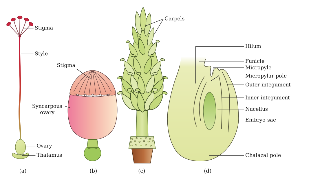
The pistil is the female reproductive unit of the flower. A flower may have a single pistil (monocarpellary) or multiple pistils (multicarpellary). When multiple pistils are present, they may be fused together (syncarpous) or remain free (apocarpous). Each pistil has three parts: the stigma, the style, and the ovary.
The stigma is the topmost part, serving as a landing platform for pollen grains. The style is the elongated, slender part beneath the stigma. The ovary is the basal, bulged part that contains the ovarian cavity (locule). Inside this cavity lies the placenta, from which the megasporangia — commonly called ovules — arise. The number of ovules in an ovary varies: some plants have a single ovule (wheat, paddy, mango), while others have many (papaya, watermelon, orchids).
Structure of the Megasporangium (Ovule)
A typical angiosperm ovule is a small structure attached to the placenta by a stalk called the funicle. The body of the ovule fuses with the funicle at a region called the hilum — the junction between the ovule and the funicle.
Each ovule has one or two protective envelopes called integuments. These integuments encircle the nucellus except at the tip, where a small opening called the micropyle is present. Opposite the micropylar end is the chalaza, which represents the basal part of the ovule.
Enclosed within the integuments is a mass of cells called the nucellus. The cells of the nucellus contain abundant reserve food materials. Located within the nucellus is the embryo sac (the female gametophyte). An ovule generally has a single embryo sac formed from a megaspore.
Megasporogenesis
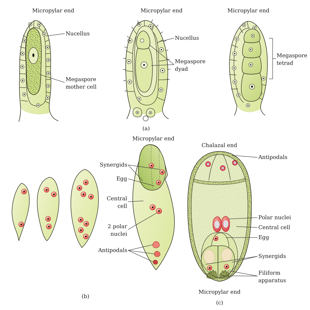
The process of formation of megaspores from the megaspore mother cell is called megasporogenesis. Ovules generally differentiate a single megaspore mother cell (MMC) in the micropylar region of the nucellus. The MMC is a large cell containing dense cytoplasm and a prominent nucleus.
The MMC undergoes meiotic division. Meiosis results in the production of four megaspores. (See the in-text question on why the MMC undergoes meiosis, below.)
Female Gametophyte (Embryo Sac)
In a majority of flowering plants, only one of the four megaspores is functional; the other three degenerate. Only the functional megaspore develops into the female gametophyte (embryo sac). This method of embryo sac formation from a single megaspore is termed monosporic development.
(See the in-text question on the ploidy of the nucellus, MMC, megaspore and female gametophyte, below.)
Formation of the Embryo Sac (Detailed)
The nucleus of the functional megaspore divides mitotically to form two nuclei, which move to opposite poles, forming the 2-nucleate embryo sac. Two more sequential mitotic nuclear divisions result in the formation of the 4-nucleate and later the 8-nucleate stages of the embryo sac.
These mitotic divisions are strictly free nuclear — nuclear divisions are not followed immediately by cell wall formation. After the 8-nucleate stage, cell walls are laid down, leading to the organisation of the typical female gametophyte (embryo sac).
Cell Distribution in the Mature Embryo Sac …
Drawn by us to help you understand the concept clearly, and verified to make sure it's accurate. For exams, practice from your textbook's own diagram.
Panels (a)-(c) compare the three ways a pistil can be built from carpels. In (a), a single carpel forms the whole pistil (monocarpellary) — Hibiscus and pea flowers work this way. In (b), several carpels are fused into one pistil sharing a single style and stigma (multicarpellary, syncarpous) — Papaver (poppy). In (c), several carpels sit side by side but stay completely separate, each with its own stigma and style, so each is technically its own tiny pistil (multicarpellary, apocarpous) — rose and lotus.
Panel (d) then opens up a single ovule — the structure inside the ovary that develops into the seed. Two protective layers, the integuments, wrap around a central tissue called the nucellus, leaving a small gap at one end called the micropyle (the opening the pollen tube will later enter through). Inside the nucellus sits the embryo sac, the female gametophyte. The ovule is anchored to the ovary wall by a stalk, the funicle, which meets the ovule body at a point called the hilum. Because the body of the ovule curves back on itself during development, the micropyle and the funicle end up close t …
Drawn by us to help you understand the concept clearly, and verified to make sure it's accurate. For exams, practice from your textbook's own diagram.
This figure follows megasporogenesis — how one diploid cell inside the ovule becomes an 8-nucleate embryo sac.
Panel (a) starts with the megaspore mother cell (MMC), a large diploid cell inside the nucellus. It undergoes meiosis I to form a dyad of two haploid cells, and each of those completes meiosis II, giving a tetrad of four haploid megaspores. In most flowering plants only one of these four megaspores stays functional (shown in teal); the other three degenerate.
Panel (b) follows the functional megaspore as its single nucleus divides mitotically without any new cell walls forming in between — first to 2 nuclei, then 4, then 8. This is why it's called free-nuclear division: at this stage the embryo sac is one large cell with 8 free nuclei floating inside it, not yet 8 separate cells. …