Physical Education · Ch 7 — Fundamentals of Anatomy and Physiology in Sports
Joints
Joints
A joint, or articulation, is the point where two or more bones meet and where muscles act on them to cause movement. Although a joint is usually thought of as movable, this is not true in every case - some joints show only limited movement and some are completely immovable.
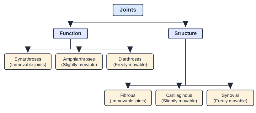
Joints are classified in two ways - by their function and by their structure.
Drawn by us to help you understand the concept clearly, and verified to make sure it's accurate. For exams, practice from your textbook's own diagram.
A flow chart headed 'Joints' that splits into two branches. The Function branch leads to Synarthroses (immovable), Amphiarthroses (slightly movable) and Diarthroses (freely movable). The Structure branch leads to Fibrous (immovable), Cartilaginous (sli …
Functional classification
The functional classification focuses on the amount of movement a joint permits:
- Synarthroses - immovable joints.
- Amphiarthroses - slightly movable joints.
- Diarthroses - freely movable joints.
The freely movable joints are found mainly in the limbs, where movement and mobility matter most. The immovable and slightly movable joints are found mostly in the axial skeleton, where the priority is protecting internal organs and making firm attachments.
Structural classification
The structural classification is based on whether fibrous tissue, cartilage or a joint cavity separates the bones:
- Fibrous joints - generally immovable.
- Cartilaginous joints - a combination of immovable and slightly movable.
- Synovial joints - freely movable.
1. Fibrous joints
In a fibrous joint the bones are united by fibrous tissue and show little or no movement. They are sub-classified by structure into sutures, syndesmoses and gomphoses:
- Sutures - a tight union that prevents any movement between the bones. Sutures are found only between the bones of the skull (cranium). The skull bones of a foetus are unfused, but after birth they slowly fuse and become fixed, making the skull immovable so that it protects the brain from impact.
- Syndesmoses - a fibrous joint in which the bones are separated by some distance and bound together by ligaments, for example the fibrous membrane connecting the distal parts of the radius and ulna. Because these joints lack flexibility, ligament injuries are common, particularly at the wrist and ankle.
- Gomphoses - a peg-in-socket joint held by ligaments. The best example is the joint between a tooth and its socket.
2. Cartilaginous joints
A cartilaginous joint unites two bones by means of cartilage and permits only very slight movement. The articulating surfaces are connected by pads (discs) of fibrocartilage. Examples include the cartilage of the growing long bones and the cartilage between the ribs and the sternum.
3. Synovial joints
Synovial joints are freely movable joints. They contain synovial fluid and are found mostly in the limbs. All synovial joints share four distinguishing features:
- Articular cartilage
- Articular capsule
- Joint cavity
- Reinforcing ligament
Types of synovial joints
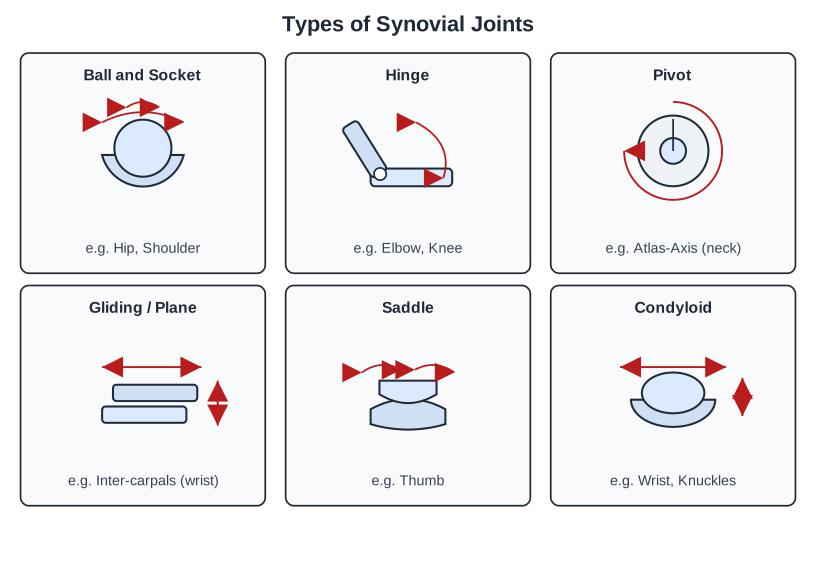
Synovial joints are classified according to the shape of the articulating surface into six types.
Drawn by us to help you understand the concept clearly, and verified to make sure it's accurate. For exams, practice from your textbook's own diagram.
A six-panel chart of the synovial joint types. Each panel has a small schematic and a label with its example: Ball and socket (hip/shoulder, movement in all axes); Hinge (elbow/knee, one-plane bending); Pivot (atlas-axis, rotation of the head); Gliding/plane (inter-carpals, sliding); Saddle (thumb, rocking t …
- Ball and socket joint. The ball-shaped head of one bone fits into the cup-like socket of another. It allows the greatest range of movement and is multiaxial - it permits movement in all axes, including rotation. Examples: the hip and shoulder joints. This joint makes possible actions such as an overhead clear in badminton or bowling in cricket.
- Hinge joint. The cylindrical end of one bone fits into a grooved surface of another, allowing a back-and-forth movement like a door hinge - that is, angular movement in one plane (flexion and extension). Examples: the elbow and knee. These movements are essential for using the biceps, triceps and quadriceps.
- Pivot joint. Also called a rotary joint, it allows only rotary movement around a single axis - the moving bone rotates within a ring formed by a second bone and its ligament. Example: the joint between the first and second cervical vertebrae (atlas and axis), which lets us turn the head from side to side. …