Biology · Ch 17 — Locomotion and Movement
Muscle
Muscle
Muscle is the tissue that gives the body most of its power to move. Before turning to muscle proper, it is worth recalling from the chapter on the cell that cilia and flagella are outgrowths of the cell membrane. Flagellar movement, produced by these whip-like extensions, is what lets spermatozoa swim, keeps water currents flowing through the canal system of sponges, and drives the locomotion of protists such as Euglena.
What muscle is
Muscle is a specialised tissue that develops from the mesoderm (the middle embryonic germ layer). It makes up a large share of the body — roughly forty to fifty per cent of the body weight of an adult human is muscle. Muscle tissue shows four special properties:
- Excitability — the ability to respond to a stimulus.
- Contractility — the ability to shorten and generate force.
- Extensibility — the ability to be stretched.
- Elasticity — the ability to return to its original shape after being stretched.
How muscles are classified
Muscles can be grouped using several different criteria: their location in the body, their appearance under the microscope, and the way their activity is regulated (whether or not it is under our conscious control). By location, three types are recognised — skeletal, visceral and cardiac.
Skeletal muscle
- Anchored directly onto the bones of the skeleton, which is exactly what lets it move them.
- A microscope reveals an alternating light-dark banding across each fibre, which is why this tissue is also known as striated muscle.
- You decide when it fires -- the conscious brain issues the command, earning it the name voluntary muscle.
- Between locomotion and everyday posture adjustments, this is the muscle type doing most of the work.
Visceral muscle
- Lines the walls of hollow internal organs -- think of the gut tube or the reproductive passages.
- No banding pattern shows up under the microscope, giving it a uniformly smooth look; hence the name smooth muscle (or nonstriated muscle).
- You have no conscious say over it firing, which is why it is labelled involuntary muscle.
- Its churning/squeezing action is what moves food along the alimentary canal and drives gametes along the genital tract.
Cardiac muscle
- Found exclusively in the wall of the heart; individual cardiac cells interconnect at branch points, knitting the whole heart muscle into one continuous, coordinated sheet.
- Even though it doesn't sit on the skeleton, it still carries the striped, banded look typical of skeletal muscle.
- Its beat is not something you consciously trigger -- an intrinsic pacemaker keeps it going, independent of deliberate nervous instruction.
The structure of a skeletal muscle
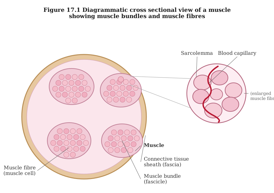
To understand how contraction works, it helps to take a skeletal muscle apart layer by layer. Each organised skeletal muscle in the body is built from several muscle bundles, also called fascicles, and these bundles are bound together by a shared covering of collagen-rich connective tissue called the fascia. Each bundle in turn holds a number of muscle fibres (the muscle cells).
Each muscle fibre has the following features:
- Its surface is bounded by a plasma membrane called the sarcolemma, which encloses the cytoplasm of the fibre, the sarcoplasm.
- A muscle fibre is a syncytium, meaning its single mass of sarcoplasm contains many nuclei.
- Its endoplasmic reticulum, here called the sarcoplasmic reticulum, acts as the store house of calcium ions.
- Running lengthwise through the sarcoplasm are large numbers of parallel filaments called myofilaments or myofibrils.
The banding pattern and the sarcomere
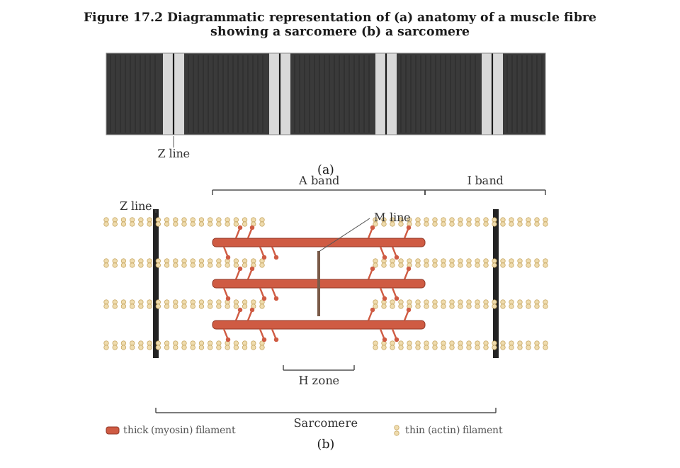
Each myofibril carries alternating dark and light bands along its length, and it is this pattern that gives striated muscle its striped look. The striations arise from the way two important proteins — actin and myosin — are laid out.
- The light band contains actin and is called the I-band or isotropic band.
- The dark band contains myosin and is called the A-band or anisotropic band.
Both proteins are arranged as rod-like structures lying parallel to one another and parallel to the long axis of the myofibril. Actin filaments are the thinner of the two and so are called thin filaments, while the stouter myosin filaments are called thick filaments.
The bands are marked out by fine reference lines: …
Drawn by us to help you understand the concept clearly, and verified to make sure it's accurate. For exams, practice from your textbook's own diagram.
This diagram cuts across a skeletal muscle to reveal how it is organised in neat layers. The whole muscle is wrapped in a sheath of connective tissue, and inside it the muscle is not one solid block but is divided into several bundles, called fascicles. One fascicle is shown enlarged so its contents are visible: it is itself made up of a cluster of long, thread-like muscle fibres, and every fibre is a single muscle cell. Each fibre's surface is bounded by a thin membrane, the sarcolemma, and running between the fibres is a blood capillary that supplies them with oxygen and nutrients. This layered packing -- muscle to bundles to fibres -- lets the muscle sta …
Drawn by us to help you understand the concept clearly, and verified to make sure it's accurate. For exams, practice from your textbook's own diagram.
This figure has two panels. Panel (a) shows the actual striped banding pattern seen along a real muscle fibre -- alternating dark and light bands repeating down its length, with a Z-line marking the boundary between one repeating unit and the next. Panel (b) diagrams that same repeating unit, the sarcomere, in schematic form: it is the stretch of myofibril lying between two Z-lines, and thousands of sarcomeres line up end to end along a myofibril. Inside each sarcomere are two kinds of filaments: thick myosin filaments and thin actin filaments that partly overlap. This overlap creates the light and dark banding you see. The I-band is light because it contains only thin filaments and is crossed by the Z-line at its centre. The A-band is dark and spans the full length of the thick filaments. In the middle of the A-band lies the H-zone, wher …