Biology · Ch 17 — Locomotion and Movement
Mechanism of Muscle Contraction
Mechanism of Muscle Contraction
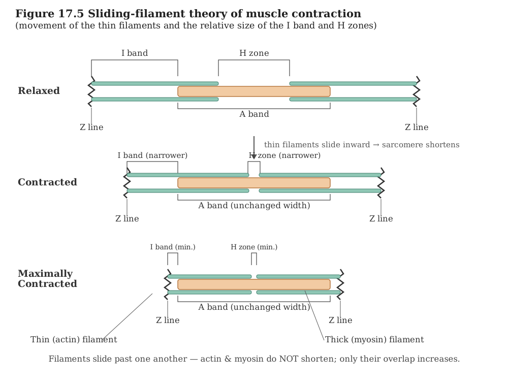
How a muscle actually shortens is explained by the sliding filament theory. Its central idea is simple: a muscle fibre contracts when the thin filaments slide over the thick filaments. The filaments themselves do not get shorter; they slide past one another so that they overlap more, and this increased overlap is what shortens the fibre.
Triggering the contraction
A contraction does not start on its own. It begins with a command from the central nervous system, which travels out along a motor neuron.
- Group a single motor neuron with every muscle fibre it innervates, and that whole set is what's termed a motor unit.
- Where that neuron actually contacts the fibre's outer membrane, the sarcolemma, is a specialised junction of its own -- the neuromuscular junction, more descriptively called the motor-end plate.
When the neural signal arrives at this junction, it causes the release of a neurotransmitter, acetylcholine. Acetylcholine generates an action potential in the sarcolemma. This action potential does not stay at one spot — it spreads through the whole muscle fibre, and as it spreads it triggers the release of calcium ions into the sarcoplasm.
The role of calcium and the exposing of active sites
The rise in the level of calcium ions is the switch that allows the filaments to interact.
- Calcium binds to a subunit of troponin, a protein sitting on the actin (thin) filaments.
- Normally the active sites on actin, the places where myosin can attach, are masked. When calcium binds troponin, this masking is removed, so the active sites on actin are exposed and become available for myosin.
Cross bridge formation and the power stroke
Once the active sites are open, the thick filaments can grip the thin filaments.
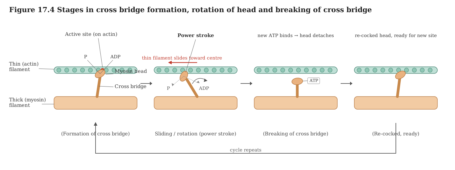
- Powered by the energy released when ATP is split, the myosin head reaches out and locks onto the now-uncovered site on actin -- this attachment is the cross bridge.
- The myosin head then rotates, and this pulls the attached actin filament towards the centre of the 'A' band.
- Because the 'Z' line is fastened to the actin filaments, it too is dragged inwards. As the Z lines on either side move closer, the sarcomere shortens — and this shortening is the contraction itself.
An important observation follows from this. During contraction the 'I' bands become reduced (their overlap increases as filaments slide together), while the 'A' bands stay the same length because the thick filaments themselves are not shortening.
The cross bridge cycle
The attachment is not permanent for a single pull. The cycle repeats to keep the filaments sliding:
- After the power stroke, the myosin head releases its ADP and inorganic phosphate and returns to its relaxed position.
- A new molecule of ATP binds to the myosin head, and this causes the cross bridge to break.
- The ATP is then hydrolysed again by the myosin head, re-energising it, and the cycle of forming a cross bridge, pulling, and breaking it repeats.
Each round of this cycle produces a little more sliding, so continued cycling gives continued shortening.
Relaxation
The muscle keeps contracting only as long as calcium is present in the sarcoplasm. Relaxation occurs when the calcium ions are pumped back into the sarcoplasmic cisternae (the stores of the sarcoplasmic reticulum). With calcium removed:
- The active sites on actin are masked once more, so myosin can no longer attach.
- The 'Z' lines return to their original positions, the sarcomere regains its resting length, and the fibre relaxes.
The reaction time, how quickly a fibre responds, is not the same in every muscle and can vary from one muscle to another.
Fatigue
When a muscle is activated again and again without adequate rest, it can become fatigued. This happens because repeated activity leads to the anaerobic breakdown of glycogen, which causes lactic acid to accumulate in the muscle. This build-up of lactic acid is what produces muscle fatigue.
Red fibres and white fibres …
Drawn by us to help you understand the concept clearly, and verified to make sure it's accurate. For exams, practice from your NCERT textbook's own diagram.
This diagram walks through the cross-bridge cycle, the repeating series of steps that actually shortens a muscle. First, when calcium exposes the active sites on actin, the energised myosin head -- still carrying the ADP and inorganic phosphate left over from splitting ATP -- attaches to actin and forms a cross bridge. Next, the head releases that ADP and phosphate as it bends or rotates, an action called the power stroke, and this pulls the thin filament a small distance towards the centre of the sarcomere. Then a fresh molecule of ATP binds to the myosin head, causing it to release its grip and detach from actin, breaking the cross bridge. Using energy from that ATP, the head re-cocks to its upright, ready position, so it can grab the actin again a little further along. As long as …
Drawn by us to help you understand the concept clearly, and verified to make sure it's accurate. For exams, practice from your NCERT textbook's own diagram.
This figure illustrates the central explanation of how a muscle shortens: the sliding-filament theory. It compares three states of the same two sarcomeres -- relaxed, contracting, and maximally contracted -- to show how the bands change as filaments slide. Going from relaxed to contracting to maximally contracted, the thin actin filaments slide progressively further inward, over the thick myosin filaments, towards the centre of each sarcomere. Because both sets of thin filaments move closer together, the Z-lines on either side are pulled nearer each other and the sarcomeres become shorter at each stage. As a result the I-band shrinks at every step, and the H-zone narrows until it nearly disappears in the maximally contracted state, while the A-band stays the same width throughout because the thick filaments do not change length. The crucial point is that neither the actin nor the myosin filaments actually shorten; they simply slide past one anoth …