Biology · Ch 4 — Principles of Inheritance and Variation
Mendelian Disorders
Mendelian Disorders
Mendelian disorders are genetic conditions caused by a mutation in a single gene. They are passed from one generation to the next in patterns that follow Mendel’s laws of inheritance — hence the name. Because they are single-gene disorders, their transmission can be traced through a family using a pedigree chart, which reveals whether the trait is dominant or recessive, and whether it is linked to an autosome or a sex chromosome.
These disorders may be dominant or recessive. Some are sex-linked (like haemophilia and colour blindness), while others are autosomal (like sickle-cell anaemia and phenylketonuria). The most common Mendelian disorders include haemophilia, cystic fibrosis, sickle-cell anaemia, colour blindness, phenylketonuria, and thalassemia.
Colour Blindness
Colour blindness is a sex-linked recessive disorder. It results from a defect in either the red or green cone cells of the eye, causing an inability to distinguish between red and green colours. The mutation occurs in certain genes present on the X chromosome.
- About 8 per cent of males and only 0.4 per cent of females are affected.
- Males have only one X chromosome, so a single recessive gene causes the disorder.
- Females have two X chromosomes; a recessive gene is usually suppressed by a dominant normal gene on the other X.
- A son of a woman who carries the gene has a 50 per cent chance of being colour blind.
- A daughter will normally be colour blind only if her mother is a carrier and her father is colour blind.
Haemophilia
Haemophilia is a sex-linked recessive disease. It is transmitted from an unaffected carrier female to some of her male progeny. The disease affects a single protein that is part of the cascade of proteins involved in blood clotting. As a result, even a simple cut can cause non-stop bleeding in an affected individual.
- A heterozygous female (carrier) may transmit the disease to her sons.
- A female becomes haemophilic only in extremely rare cases — her mother must be at least a carrier, and her father must be haemophilic (which is often unviable in later life).
- The family pedigree of Queen Victoria shows a number of haemophilic descendants, as she was a carrier.
Sickle-Cell Anaemia
Sickle-cell anaemia is an autosome-linked recessive trait. It is transmitted from parents to offspring only when both partners are carriers (heterozygous) for the gene.
- The disease is controlled by a single pair of alleles: HbA and HbS.
- Only individuals homozygous for HbS (HbS HbS) show the diseased phenotype.
- Heterozygous individuals (HbA HbS) appear unaffected but are carriers — there is a 50 per cent probability of transmitting the mutant gene to progeny. This condition is called the sickle-cell trait.
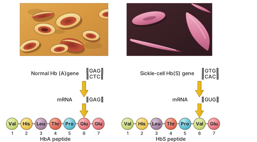
Molecular basis: The defect is caused by the substitution of the amino acid glutamic acid (Glu) by valine (Val) at the sixth position of the beta globin chain of haemoglobin. This substitution results from a single base change at the sixth codon of the beta globin gene — from GAG to GUG.
Effect: Under low oxygen tension, the mutant haemoglobin molecules undergo polymerisation. This changes the shape of red blood cells from a biconcave disc to an elongated, sickle-like structure.
Phenylketonuria
Phenylketonuria is an inborn error of metabolism inherited as an autosomal recessive trait.
- The affected individual lacks the enzyme that converts the amino acid phenylalanine into tyrosine.
- As a result, phenylalanine accumulates and is converted into phenylpyruvic acid and other derivatives.
- Accumulation of these substances in the brain causes mental retardation.
- These derivatives are also excreted through urine because of poor absorption by the kidney.
Thalassemia
Thalassemia is an autosome-linked recessive blood disease. It is transmitted from parents to offspring when both partners are unaffected carriers (heterozygous) for the gene.
- The defect is due to either mutation or deletion, which results in a reduced rate of synthesis of one of the globin chains (alpha or beta) that make up haemoglobin.
- This leads to the formation of abnormal haemoglobin molecules, causing anaemia — the characteristic feature of the disease.
Classification: …
Drawn by us to help you understand the concept clearly, and verified to make sure it's accurate. For exams, practice from your NCERT textbook's own diagram.
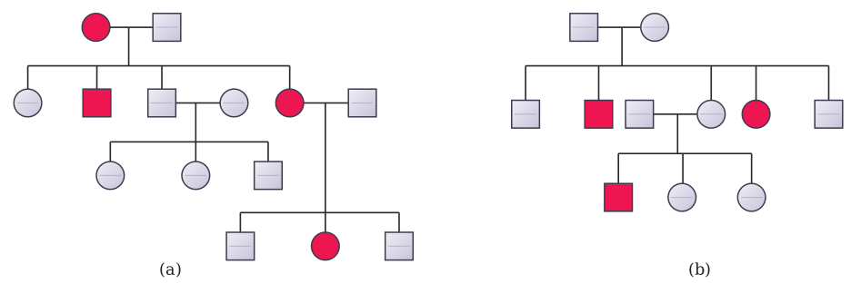
Figure 4.14 presents two separate pedigree charts side by side, labelled (a) and (b). Each chart uses the standard pedigree symbols: squares for males, circles for females, a filled (shaded) symbol for an affected individual, and an unfilled symbol for an unaffected individual. Horizontal lines connect parents, and vertical lines lead down to their children, arranged in rows by generation (typically Roman numerals I, II, III, etc., though the figure itself may not show generation labels explicitly — the pattern is what matters).
Panel (a) shows the inheritance of an autosomal dominant trait, using myotonic dystrophy as the example. The key feature is that the trait appears in every generation: at least one affected parent in each generation has affected children. You will see an affected parent (say, an affected father in generation I) passing the trait directly to some of his children in generation II — both sons and daughters are affected, because the gene is on an autosome, not a sex chromosome. Those affected children, in turn, have affected offspring in generation III. No unaffected parents produce an affected child, because a dominant allele cannot be hidden — if a child has the trait, one parent must also have it. The pattern is vertical: the trait does not skip generations.
Panel (b) shows the inheritance of an autosomal recessive trait, using sickle-cell anaemia as the example. Here the trait can skip generations. Typically, you see unaffected parents (both carriers, shown as unfilled symbols, though carriers are not always marked differently in a basic pedigree — the textbook often uses a half-filled symbol or a dot, but the figure as described uses only filled/unfilled). The key is that two unaffected parents produce an affected child (filled symbol) in the next generation. That child must be homozygous recessive. The parents are both heterozygous carriers. The trait may appear in siblings but not in the parents or grandparents — it appears to "skip" a generation. When an affected individual has children, they will pass the recessive allele to all their offspring, but unless the other parent is also a carrier or affected, the children will be unaffected carriers. So the pattern is horizontal: affected individuals often appear in a single generation among siblings, with no affected parent. …
Drawn by us to help you understand the concept clearly, and verified to make sure it's accurate. For exams, practice from your NCERT textbook's own diagram.
Figure 4.15 is a composite image that pairs a micrograph of red blood cells with a diagram of the amino acid sequence of the beta-globin chain of haemoglobin. The figure is split into two panels, (a) and (b), placed side by side for direct comparison.
Panel (a) shows the normal condition. The micrograph displays red blood cells that are typical biconcave discs — round, indented in the centre, and uniform in shape. Beside this micrograph, a short segment of the beta-globin chain is shown, with the amino acid at the sixth position labelled as glutamic acid (Glu). The corresponding codon in the beta-globin gene is GAG.
Panel (b) shows the condition in sickle-cell anaemia. The micrograph here reveals red blood cells that have lost their normal disc shape and are elongated, curved, and pointed — resembling a sickle or crescent. Beside this micrograph, the same segment of the beta-globin chain is depicted, but now the sixth position carries valine (Val) instead of glutamic acid. The codon change is shown as GUG (a single base substitution from GAG to GUG).
The figure uses arrows or a direct visual alignment to connect the change in the amino acid sequence to the change in red blood cell shape. The message is clear: a single nucleotide substitution in the beta-globin gene leads to a single amino acid substitution (Glu → Val) at the sixth position of the beta chain. This altered haemoglobin (HbS) polymerises under low oxygen tension, distorting the red blood cell from a flexible biconcave disc into a rigid sickle shape.
The figure illustrates the molecular basis of an autosomal recessive Mendelian disorder: a point mutation (GAG → GUG) in the beta-globin gene causes a glutamic acid to valine substitution, which directly produces the sickled RBC phenotype seen in homozygous individuals (HbS HbS). …