Biology · Ch 1 — Sexual Reproduction in Flowering Plants
Double Fertilisation
Double Fertilisation
What Double Fertilisation Is
After the pollen tube enters one of the synergids, it releases its two male gametes into the synergid's cytoplasm. From there, two separate fusion events take place inside the same embryo sac: one male gamete fuses with the egg cell, and the other fuses with the two polar nuclei of the central cell. This process is called double fertilisation, and it is unique to angiosperms (flowering plants) — no other group of plants or animals does this.
What Happens During Double Fertilisation
The journey begins when a pollen grain lands on a compatible stigma and germinates. The pollen tube grows down through the style, carrying two male gametes (sperm cells) inside it. The tube enters the embryo sac through the micropyle — the small opening at the base of the ovule.
Inside the embryo sac, the tip of the pollen tube ruptures, releasing the two sperm cells. Now the critical events unfold:
-
One sperm fuses with the egg cell (the female gamete). This fusion produces a diploid zygote (2n). This is syngamy — the true fertilisation that gives rise to the embryo.
-
The other sperm fuses with the two polar nuclei (which are already present in the central cell of the embryo sac). This triple fusion produces a triploid primary endosperm nucleus (3n). This is the second fertilisation event.
Because two fertilisation events occur inside the same embryo sac, the entire process is called double fertilisation.
The two polar nuclei may fuse before the sperm arrives, forming a single diploid secondary nucleus. In that case, the sperm fuses with this already-formed secondary nucleus. Either way, the result is a triploid primary endosperm nucleus.
The Two Products of Double Fertilisation
| Fertilisation event | Cells involved | Product | Ploidy | Fate |
|---|---|---|---|---|
| Syngamy (first) | Sperm + egg | Zygote | 2n (diploid) | Develops into the embryo |
| Triple fusion (second) | Sperm + two polar nuclei | Primary endosperm nucleus | 3n (triploid) | Develops into the endosperm (nutritive tissue for the embryo) |
The primary endosperm nucleus divides repeatedly to form the endosperm, a triploid tissue that stores food (starch, proteins, oils) for the developing embryo. In many seeds, the endosperm persists and is consumed by the embryo during germination (e.g., in cereals like wheat and maize). In others, the endosperm is absorbed by the cotyledons before the seed matures (e.g., in peas and beans).
Why Double Fertilisation Matters
Double fertilisation ensures that endosperm development is triggered only after successful fertilisation. If no fertilisation occurs, no endosperm forms — the ovule simply aborts. This is an efficient system: the plant does not waste resources building nutritive tissue for an unfertilised ovule.
The triploid nature of the endosperm also provides a genetic advantage. Because it has two maternal sets of chromosomes and one paternal set, the endosperm can support vigorous embryo growth while maintaining a balance between maternal and paternal contributions. …
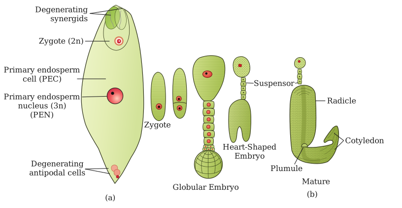
Drawn by us to help you understand the concept clearly, and verified to make sure it's accurate. For exams, practice from your NCERT textbook's own diagram.
Figure 1.13 is split into two panels, (a) and (b), that together tell the story of what happens inside the ovule after double fertilisation.
Panel (a) shows a single, fertilised embryo sac. The sac is drawn as an elongated oval, with the micropylar end (the entry point for the pollen tube) at the bottom and the chalazal end at the top. Inside, you see the key products of the two fusion events. At the micropylar end, near where the egg cell used to be, sits the zygote — a single diploid cell formed by syngamy. In the centre of the sac, occupying the large central cell, is the Primary Endosperm Nucleus (PEN) — a triploid nucleus formed by triple fusion (the fusion of one male gamete with the two polar nuclei). The synergids and antipodals are not shown; the focus is entirely on these two cells that will give rise to the next generation. The zygote and PEN are the only labelled structures in this panel, and their positions (zygote at the micropylar end, PEN in the central cell) are critical for understanding what happens next.
Panel (b) is drawn at a smaller scale than (a), as the caption notes. It shows a sequence of stages in embryo and endosperm development in a dicot plant, arranged from left to right. The first stage shows the proembryo — a small, undifferentiated cluster of cells derived from the zygote, still attached to the suspensor (a short chain of cells that anchors the embryo and pushes it into the endosperm). Next comes the globular stage, where the embryo is a rounded mass of cells. Then the heart-shaped stage, where the embryo begins to show bilateral symmetry — two cotyledon primordia bulge out on either side, giving it a heart-like outline. Throughout these stages, the surrounding endosperm is shown as a mass of cells (or free nuclei, depending on the stage) that fills the embryo sac and provides nutrition to the developing embryo. The endosperm develops first, as the textbook explains, and the embryo grows into it.
The relationship between the two panels is sequential: (a) shows the starting point (zygote + PEN), and (b) shows the developmental outcome (embryo stages + endosperm). The arrows or progression in (b) indicate that the zygote divides repeatedly to form the proembryo, then the globular and heart-shaped stages, while the PEN divides to form the endosperm tissue that surrounds and nourishes the embryo. No other labels (like suspensor, cotyledons, or radicle) are mentioned in the description, so the figure keeps the focus on the broad sequence rather than detailed anatomy. …