Biology · Ch 7 — Human Health and Disease
Immune System in the Body
Immune System in the Body
The immune system is not a single organ but a distributed network. It includes lymphoid organs, tissues, cells, and soluble molecules like antibodies. What makes it unique is its ability to recognise foreign antigens, respond to them, and then remember them for future encounters. Beyond fighting infections, the immune system is also central to allergic reactions, auto-immune diseases, and the success or failure of organ transplants.
Lymphoid Organs
Lymphoid organs are the sites where lymphocytes originate, mature, and proliferate. They are divided into two categories based on their function.
Primary lymphoid organs are where immature lymphocytes develop into antigen-sensitive lymphocytes. There are two:
- Bone marrow: The main lymphoid organ. It produces all blood cells, including lymphocytes.
- Thymus: A lobed organ located near the heart, beneath the breastbone. It is quite large at birth but shrinks with age, becoming very small by puberty. Both bone marrow and the thymus provide the micro-environment needed for the development and maturation of T-lymphocytes.
After maturation, lymphocytes migrate to secondary lymphoid organs. These organs provide the sites where lymphocytes actually encounter antigens, interact with them, and then proliferate to become effector cells. The secondary lymphoid organs include:
- Spleen: A large, bean-shaped organ. It contains mainly lymphocytes and phagocytes. It acts as a blood filter, trapping blood-borne micro-organisms. It also serves as a large reservoir of erythrocytes (red blood cells).
- Lymph nodes: Small, solid structures located at different points along the lymphatic system. They trap micro-organisms or other antigens that enter the lymph and tissue fluid. The trapped antigens activate the lymphocytes present in the nodes, triggering an immune response.
- Tonsils, Peyer’s patches of the small intestine, and the appendix are also secondary lymphoid organs. …
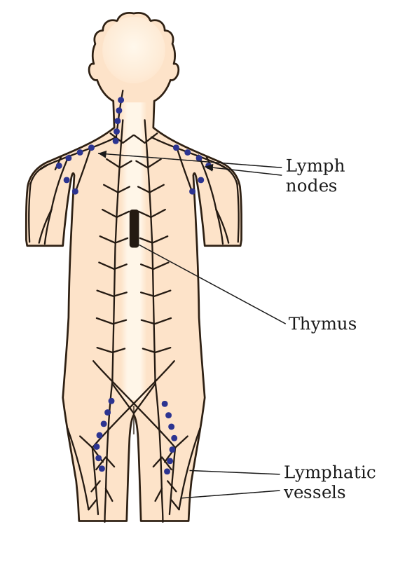
Drawn by us to help you understand the concept clearly, and verified to make sure it's accurate. For exams, practice from your textbook's own diagram.
The figure marks the location of lymphoid tissue on an outline of the human body. The real NCERT figure (and this platform's own SVG) labels only three things: lymph nodes, the thymus, and lymphatic vessels — it does not show bone marrow, the spleen, tonsils, Peyer's patches, or the appendix.
Lymph nodes are shown as small clusters at several points along the body's lymphatic pathways — in the neck (cervical nodes), armpits (axillary nodes), and groin (inguinal nodes) — connected by a network of lymphatic vessels that drain fluid from the tissues. The thymus is shown as a small lobed gland just below the neck, behind the breastbone; it is the site where T-lymphocytes mature, and is notably large at birth but shrinks by puberty. …