Biology · Ch 5 — Molecular Basis of Inheritance
Packaging of DNA Helix
Packaging of DNA Helix
The DNA in a typical mammalian cell is astonishingly long. If you take the distance between two consecutive base pairs as 0.34 nm (that is 0.34 × 10⁻⁹ m) and multiply it by the total number of base pairs — roughly 6.6 × 10⁹ bp — the total length of the DNA double helix comes out to about 2.2 metres. Yet this entire length is packed inside a nucleus that is only about 10⁻⁶ m across. How is that possible?
The answer lies in a highly organised packaging system, and it works differently in prokaryotes and eukaryotes.
Packaging in Prokaryotes
Prokaryotes like E. coli do not have a defined nucleus, but their DNA is not scattered randomly. The DNA is negatively charged (due to the phosphate backbone), and it is held together with positively charged proteins in a region called the nucleoid. Within the nucleoid, the DNA is organised into large loops that are held in place by these proteins. (For reference, the length of E. coli DNA is 1.36 mm; you can calculate the number of base pairs by dividing that length by 0.34 nm per bp.)
Packaging in Eukaryotes
Eukaryotic packaging is far more complex. The key players are a set of positively charged, basic proteins called histones. A protein's charge depends on the abundance of amino acid residues with charged side chains. Histones are rich in the basic amino acids lysine and arginine, both of which carry positive charges in their side chains.
Histones organise themselves into a unit of eight molecules called a histone octamer. The negatively charged DNA wraps around this positively charged octamer to form a structure called a nucleosome. A typical nucleosome contains about 200 base pairs of DNA.
Nucleosomes are the repeating unit of a larger structure in the nucleus called chromatin — the thread-like, stained bodies you see in the nucleus. When viewed under an electron microscope, nucleosomes in chromatin look like 'beads-on-a-string'. (Theoretically, in a mammalian cell with 6.6 × 10⁹ bp of DNA, and each nucleosome containing 200 bp, you would have roughly 3.3 × 10⁷ such beads.) …
Drawn by us to help you understand the concept clearly, and verified to make sure it's accurate. For exams, practice from your textbook's own diagram.
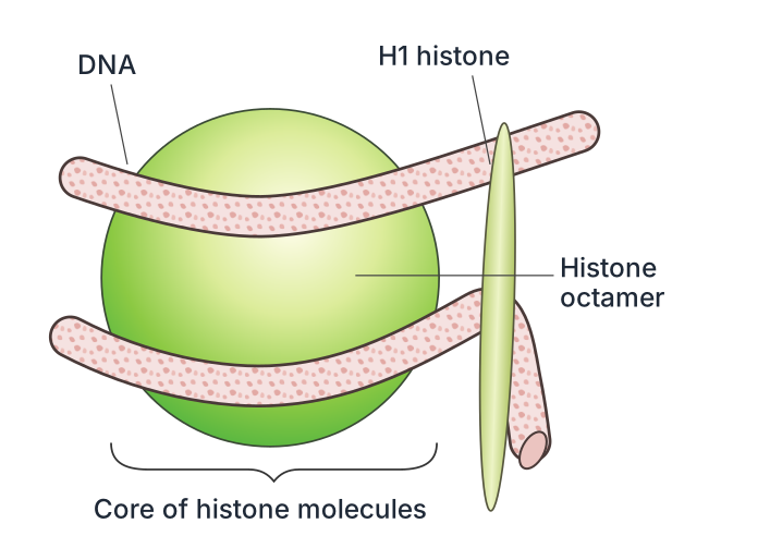
Figure 5.4a is a schematic diagram of a single nucleosome — the fundamental repeating unit of eukaryotic chromatin. The drawing shows a central, roughly cylindrical core made of eight histone proteins (the histone octamer), around which DNA is wound. The DNA is depicted as a continuous double helix that makes about 1.7 turns around the octamer, entering and exiting the structure from opposite sides. The two ends of the DNA segment are shown as free, short stretches that lead to the next nucleosome in the chromatin fiber.
The histone octamer itself is not drawn as eight separate blobs; instead, it is represented as a single compact, bead-like shape, with the DNA wrapped tightly around its surface. The figure does not label individual histone proteins (H2A, H2B, H3, H4) — it simply shows the octamer as a unit. The DNA is labelled as "DNA" along its length, and the entire assembly is labelled "Nucleosome". There are no arrows, no colour coding, and no additional panels in this part of the figure. The key relationship it teaches is that the negatively charged DNA backbone is electrostatically attracted to the positively charged histone octamer, allowing the long DNA molecule to be spooled into a compact, repeating structure.
The figure does not show the linker DNA between nucleosomes, the H1 histone, or the "beads-on-string" appearance — those are depicted in Figure 5.4b. Here, the focus is solely on the nucleosome core particle itself. …
Drawn by us to help you understand the concept clearly, and verified to make sure it's accurate. For exams, practice from your textbook's own diagram.
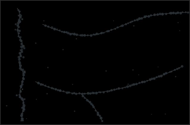
The figure is an electron micrograph — a black-and-white image taken through an electron microscope — showing a single, thin chromatin fibre stretched out. Along the length of this fibre you see a repeating pattern: small, roughly spherical dark blobs spaced at regular intervals, connected by very thin, faintly visible threads of DNA. This is the classic "beads-on-a-string" appearance.
Each dark blob is a nucleosome: the fundamental packaging unit of eukaryotic chromatin. The "string" between them is the linker DNA — a short stretch of double-helical DNA that runs from one nucleosome to the next. The nucleosome itself consists of a histone octamer (a core of eight positively charged histone proteins) around which the negatively charged DNA is wrapped approximately 1.65 turns, covering about 146 base pairs. The textbook states that a typical nucleosome contains 200 bp of DNA — this includes the wrapped portion plus the linker DNA that connects to the next bead.
The image teaches a key lesson in molecular packing: the DNA in a single human cell is about 2 metres long, yet the nucleus is only a few micrometres across. The first level of compaction is this nucleosomal arrangement. By winding DNA around histones, the linear length is reduced roughly sevenfold — from a continuous double helix to a compact, repeating chain of beads. This "beads-on-string" structure is the 10 nm chromatin fibre, the most basic level of chromatin organisation. It is not the final form; in the cell, these beads are further coiled and folded into thicker fibres (30 nm and beyond), eventually condensing into metaphase chromosomes. But the figure captures the very first, essential step: how the long, thin DNA molecule is systematically spooled onto protein cores to fit inside the nucleus. …