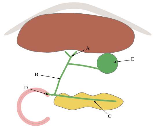
Q.Label the given diagram.
A
B
C
D
E
You're viewing a preview — the full solution, concept, methods & PYQ mapping are locked.
Start your 14-day free trial to unlock the full solution →Step 1. Identify the organs by shape and colour. The large reddish-brown organ at the top is the liver; the yellow lobulated gland beside the curved pink tube at the bottom is the pancreas; the pink coiled tube is the duodenum (first part of the small intestine); the small green pouch is the gallbladder — label E points directly at it.
Step 2. Trace the duct leaving the liver (A). Bile produced continuously by liver cells drains into small ducts that unite to form the (common) hepatic duct, which carries bile away from the liver — this is what label A marks, at the point just below the liver where the duct begins.
Step 3. Follow the duct to B. The hepatic duct is joined by the cystic duct coming from the gallbladder (which stores and concentrates bile between meals). Once these two ducts unite, the single duct that continues downward is called the common bile duct — this is the duct marked B.
Step 4. Identify the pancreatic contribution (C). Running through/alongside the pancreas is the pancreatic duct, which collects the digestive enzyme-rich pancreatic juice from the pancreatic acini — this is what label C marks. …
Unlock everything free for 14 days
- Full step-by-step solutions
- Concept-first explanations
- Methods, shortcuts & mistakes
- PYQ mapping + timed mock tests
Full access for 14 days. No credit card required.