Q.Describe the internal structure of a monocot root.
You're viewing a preview — the full solution, concept, methods & PYQ mapping are locked.
Start your 14-day free trial to unlock the full solution →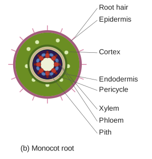
A monocot root shows epiblema → cortex → endodermis → pericycle → radial, polyarch vascular bundles surrounding a large, well-developed pith; it lacks cambium, so it never undergoes secondary growth.
A transverse section (T.S.) of a typical monocotyledonous root, taken through the maturation/piliferous region, shows the following tissues arranged from outside to inside:
-
Epiblema (epidermis): The outermost, single layer of thin-walled cells, without a cuticle, forming unicellular root hairs that increase the absorptive surface area for water and mineral uptake.
-
Cortex: A wide zone of thin-walled, parenchymatous cells with numerous intercellular spaces, lying just beneath the epiblema; functions in storage and lateral movement of water.
-
Endodermis: The innermost layer of the cortex, a single layer of barrel-shaped cells bearing casparian strips (band-like thickenings of suberin) on their radial and inner tangential walls, regulating the movement of water/minerals into the vascular tissue. Some thin-walled cells opposite the protoxylem, called passage cells, lack the casparian strip thickening and allow radial water movement.
-
Pericycle: A single layer of cells just inside the endodermis, from which lateral roots and the vascular cambium (in dicots) arise; in monocots it retains a purely meristematic role for lateral root initiation since no cambium develops.
…
Unlock everything free for 14 days
- Full step-by-step solutions
- Concept-first explanations
- Methods, shortcuts & mistakes
- PYQ mapping + timed mock tests
Full access for 14 days. No credit card required.