Botany · Ch 2 — Biological Classification
Eubacteria
Eubacteria
There are thousands of different eubacteria, or 'true bacteria'. They are characterised by the presence of a rigid cell wall, and, if motile, a flagellum for movement.
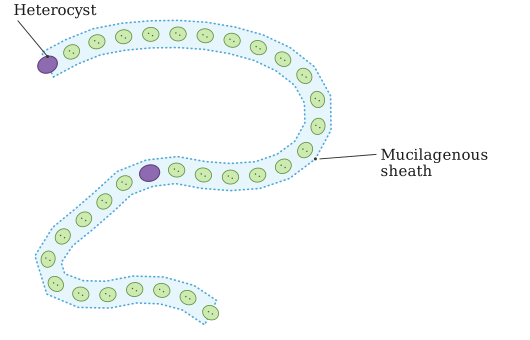
The cyanobacteria, also referred to as blue-green algae, are an important group of eubacteria. They contain the pigment chlorophyll a, similar to green plants, and are photosynthetic autotrophs. Cyanobacteria may be unicellular, colonial or filamentous, and are found in freshwater, marine or terrestrial habitats. The colonies are generally surrounded by a gelatinous sheath, and these organisms often form blooms in polluted water bodies. Some cyanobacteria can fix atmospheric nitrogen in specialised cells called heterocysts — for example, Nostoc and Anabaena.
Chemosynthetic autotrophic bacteria oxidise various inorganic substances such as nitrates, nitrites and ammonia, and use the released energy for their ATP production. They play a great role in recycling nutrients like nitrogen, phosphorous, iron and sulphur. …
Drawn by us to help you understand the concept clearly, and verified to make sure it's accurate. For exams, practice from your textbook's own diagram.
This figure illustrates Nostoc, a filamentous cyanobacterium (blue-green algae) belonging to the Eubacteria. The organism is drawn as a chain of rounded cells joined end to end to form a long, beaded filament. The whole filament is enclosed within a mucilagenous sheath, the gelatinous covering typical of cyanobacterial colonies. At intervals along the chain, larger, specialised cells called heterocysts are marked; these are the sites where Nostoc can fix atmospheric nitrogen. Being photosynthetic autotrophs that contain chlorophyll a, such cyanobacteria often fo …
Drawn by us to help you understand the concept clearly, and verified to make sure it's accurate. For exams, practice from your textbook's own diagram.
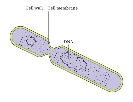
This figure shows a bacterium in the act of reproducing by fission, the main mode of reproduction in Monera. The rod-shaped cell is caught midway through splitting into two daughter cells of roughly equal size. The drawing labels the cell wall, the rigid outer covering that gives the bacterium its shape, and the cell membrane lying just inside it. Within the cytoplasm the DNA is shown as a coiled mass; because bacteria are prokaryotic, this genetic material is not enclosed in a nuclear membrane. During fission the DNA is copied and the cel …