Zoology · Ch 2 — Structural Organisation in Animals
Anatomy
Anatomy
The anatomy of the frog reveals several well-developed organ systems, each with a clear division of labour.
-
Digestive system. The alimentary canal runs from the mouth through the buccal cavity, pharynx and oesophagus into the stomach, then the intestine (whose first part is the duodenum) and the short rectum, ending in the cloaca — a common chamber that also receives the urinary and reproductive products and opens out through the cloacal aperture. Because frogs are carnivorous, the intestine is comparatively short. Digestive glands include the liver (which secretes bile stored in the gall bladder) and the pancreas (which secretes pancreatic juice with digestive enzymes). Food is captured by the bilobed tongue; digestion begins in the stomach with HCl and gastric juice, which reduce the food to a semi-digested mass called chyme. Chyme passes into the duodenum, the first part of the intestine, which receives bile from the gall bladder and pancreatic juice from the pancreas through a shared common bile duct — bile emulsifies fats while pancreatic juice digests carbohydrates and proteins, with digestion finishing in the intestine. Digested food is absorbed through the numerous finger-like villi and microvilli lining the intestine's inner wall, and undigested matter is expelled through the cloaca.
-
Respiratory system. A frog respires in more than one way. On land it mainly uses its lungs (pulmonary respiration) — a pair of elongated, pinkish, elastic sac-like structures in the upper part of the trunk (thorax); air travels in through the nostrils, across the buccal cavity, and into the lungs. In water, and while otherwise inactive, gaseous exchange happens across the moist, richly vascularised skin (cutaneous respiration) by simple diffusion of dissolved oxygen; the lining of the buccal cavity also participates in gas exchange. During aestivation and hibernation, when the frog is dormant, respiration occurs through the skin alone.
-
Circulatory system. The frog has a closed blood vascular system with a three-chambered heart — two atria and a single ventricle — enclosed in a pericardium. A triangular chamber called the sinus venosus opens into the right atrium and receives blood from the great veins, the venae cavae; on the ventral side, the ventricle in turn opens into a sac-like conus arteriosus, from which blood is carried onward to the body through the arterial system, while the venous system returns it from the body to the heart. Special venous connections — the hepatic portal system (linking the intestine and liver) and the renal portal system (linking the kidney and the lower body) — are also present. Alongside the blood vascular system there is a lymphatic system of lymph, lymph channels and lymph nodes. Blood itself is made of plasma and cells — RBCs (erythrocytes), which are nucleated and carry the red respiratory pigment haemoglobin, WBCs (leucocytes), and platelets — and it carries nutrients, gases and water to the tissues.
-
Excretory system. It consists of a pair of kidneys, the ureters, the urinary bladder and the cloaca. The kidneys filter nitrogenous wastes from the blood; the ureters carry the urine to the cloaca, from where it is passed out. The frog is ureotelic, excreting mainly urea.
-
Nervous and sensory systems. The nervous system is organised into a central part (brain and spinal cord), a peripheral part — ten pairs of cranial nerves arising directly from the brain, plus the spinal nerves — and an autonomic part (sympathetic and parasympathetic). The brain is enclosed in a bony brain-box, the cranium, and is divided into three regions: a forebrain, made up of the olfactory lobes, the paired cerebral hemispheres and the unpaired diencephalon; a midbrain, marked by a pair of optic lobes; and a hindbrain, made up of the cerebellum and the medulla oblongata. The medulla oblongata leaves the skull through an opening called the foramen magnum and continues on as the spinal cord, which runs enclosed within the vertebral column. The frog's sense organs cover touch (sensory papillae), taste (taste buds), smell (the nasal epithelium), sight (the eyes) and hearing (the tympanum, together with the internal ear); of these, only the eyes and internal ears are well-organised structures, the rest being simpler clusters of cells around nerve endings. The eyes are a pair of simple, single-unit spherical structures set in the skull's orbits; frogs have no external ear at all — only the externally visible tympanum, which serves both hearing and balance (equilibrium). …
Drawn by us to help you understand the concept clearly, and verified to make sure it's accurate. For exams, practice from your textbook's own diagram.
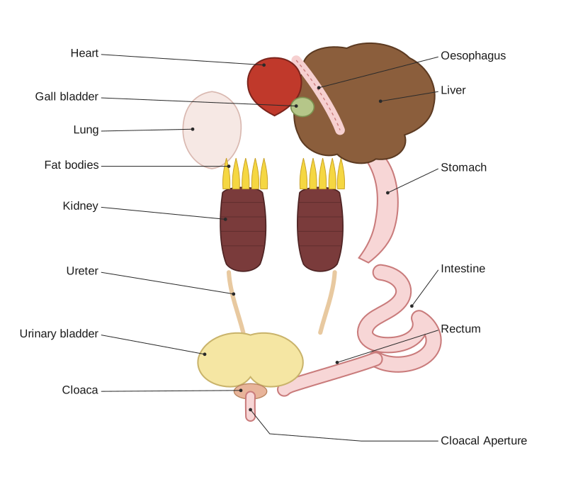
This dissection-style diagram exposes the body cavity of the frog to show its internal organs arranged around the alimentary canal. At the top the three-chambered heart lies in the midline, with the oesophagus and the large brown liver flanking it and the small gall bladder tucked beneath. On the right the tube continues into the stomach and then into the coiled intestine, which narrows into the rectum before opening at the cloaca and cloacal aperture at the bottom. On the left the paired lungs, the yellow finger-like fat bodies, the dark bean-shaped kidneys, the ureters, the urinary bladder and the cloaca are labelled, so the figure toge …
Drawn by us to help you understand the concept clearly, and verified to make sure it's accurate. For exams, practice from your textbook's own diagram.
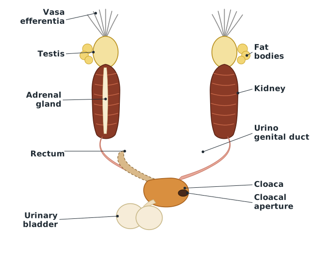
This figure shows the urinogenital organs of a male frog as a paired, roughly symmetrical arrangement lying against the dorsal body wall. The two yellowish, ovoid testes are drawn adhered to the upper part of the kidneys; fine tubules called vasa efferentia, 10 to 12 in number, are shown arising from each testis and entering the kidney on its own side. Above the kidneys sit the yellow, branched fat bodies, and the small adrenal gland is marked on the kidney surface. From each kidney a urinogenital duct runs downward, because in the male the same duct carries both urine and sperm. The ducts, together with the **rec …
Drawn by us to help you understand the concept clearly, and verified to make sure it's accurate. For exams, practice from your textbook's own diagram.
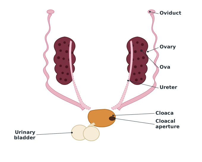
This figure shows the reproductive and associated organs of a female frog. The most prominent structures are the two large, dark, lobed ovaries lying in the body cavity; their surface is studded with numerous rounded ova, illustrating that a mature female can carry and release 2500 to 3000 eggs at a time. Arising near the ovaries are the paired, coiled oviducts, drawn as long convoluted tubes that carry the released ova; unlike the male, the female has no functional connection between the ovaries and the kidneys. The ureters are shown separately draining the kidneys. Below, the oviducts, ureters and the **urinary …