Question 84 of 91
Q.Draw a neat and labelled diagram of Ommatidium.
Telangana TsbieTelangana Board of Intermediate Education (Intermediate 1st Year) 2020Subjective· 4mImportance★★★★★
92% · 84/91 Questions
You're viewing a preview — the full solution, concept, methods & PYQ mapping are locked.
Start your 14-day free trial to unlock the full solution →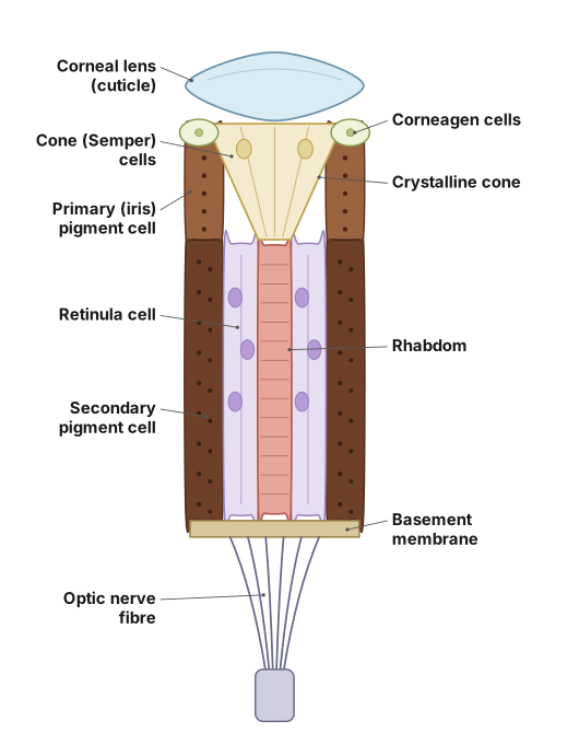
An ommatidium is the single structural/functional unit of the compound eye; thousands of them together form the compound eye of the cockroach.
Note on the figure: This question asks for a drawn, labelled diagram. A hand-drawn image cannot be produced in this text answer, so instead the structure is described part-by-part, in the same order they would be labelled top-to-bottom on a diagram, so the layout can be reconstructed and drawn accurately.
Structure of an ommatidium (outer to inner):
- Cuticle/Corneal lens: the outermost transparent, biconvex cuticular lens that admits and focuses light.
- Corneagen cells: a pair of cells beneath the cornea that secrete/renew the corneal lens.
- Crystalline cone: a transparent, cone-shaped structure just beneath the cornea that further focuses light onto the light-sensitive part below; surrounded by four cone cells (Semper cells).
- Primary (iris) pigment cells: surround the crystalline cone, optically isolating each ommatidium from its neighbours.
- Retinula cells: a group of (typically 7-8) elongated sensory (photoreceptor) cells arranged radially around a central axis, beneath the crystalline cone.
- Rhabdom: a central, rod-like structure formed by the fused inner (microvillar) borders of the retinula cells; it is the actual light-sensitive photoreceptive element that converts light into nerve impulses. …
Unlock everything free for 14 days
- Full step-by-step solutions
- Concept-first explanations
- Methods, shortcuts & mistakes
- PYQ mapping + timed mock tests
Full access for 14 days. No credit card required.