Q.Which of the following enzymes catalyse the removal of nucleotides from the ends of DNA?
Concept understanding — Restriction Enzyme Action
Imagine you have a long, tangled piece of string, and you need to cut it into smaller, specific pieces — not just anywhere, but exactly at the places where a certain pattern of letters appears. That is the core idea behind restriction enzyme action.
In the world of biology, the "string" is a DNA molecule — the long, thread-like chemical that carries the genetic instructions for every living thing. A restriction enzyme is a molecular "scissors" that cuts DNA, but it is incredibly precise. It does not chop randomly. Instead, it recognises a very specific, short sequence of DNA letters (usually 4 to 8 base pairs long) and cuts only at that exact spot.
Think of it like a word processor's "Find and Replace" function, but instead of replacing text, the enzyme finds a specific word and cuts the page at that word.
This ability to cut DNA at precise locations is what makes restriction enzymes the fundamental tool of genetic engineering. Without them, scientists would have no way to isolate a specific gene from a long DNA strand.
How does the enzyme "know" where to cut?
The DNA molecule is made of two strands twisted together (the famous double helix). Each strand has a sequence of four chemical "letters": A, T, G, and C. A restriction enzyme scans along the DNA until it finds its target sequence — a short, palindromic pattern (meaning it reads the same forwards on one strand and backwards on the other). For example, the enzyme EcoRI recognises the sequence GAATTC.
When it finds this exact sequence, it binds to the DNA and makes a cut in both strands. The cut can be one of two types:
- Sticky ends: The enzyme cuts the two DNA strands at different points, leaving short, single-stranded overhangs. These overhangs are like pieces of Velcro — they can easily stick to a complementary overhang from another DNA piece cut by the same enzyme. This is extremely useful for joining different DNA fragments together.
- Blunt ends: The enzyme cuts both strands straight across at the same point, leaving no overhang. These are harder to join together later, but they are still useful.
Why does this matter?
Restriction enzymes are the reason we can manipulate DNA at all. They allow scientists to:
- Cut out a specific gene from the DNA of one organism (say, the human insulin gene).
- Cut open a carrier DNA (like a plasmid from a bacterium) at the same spot.
- Insert the gene into the carrier, because the sticky ends match perfectly.
- Splice the carrier back together using another enzyme (DNA ligase), creating a recombinant DNA molecule.
This is the foundation of modern biotechnology — from producing human insulin in bacteria to creating genetically modified crops and developing gene therapies.
The key takeaway from the NCERT textbook is that restriction enzymes are molecular scissors that cut DNA at specific recognition sites. Their action produces fragments with either sticky ends or blunt ends, and this precise cutting is what makes genetic engineering possible. The enzyme itself is a protein, and it is named after the bacterium from which it is isolated (e.g., EcoRI from Escherichia coli).
In short: Restriction enzyme action is the controlled, precise cutting of DNA at predetermined locations — the first and most essential step in any DNA manipulation experiment.
Restriction enzyme action is one of the most tested mechanisms in the NCERT Class 12 Biology chapter on Biotechnology: Principles and Processes, and appears in searches like "restriction enzymes class 12 biology sticky ends" or "EcoRI recognition site important questions." This is a near-certain topic in both CBSE board papers and NEET's biotechnology section every year.
Nucleases are enzymes that cleave the phosphodiester bonds of nucleic acids. They are broadly classified based on their site of action on the DNA molecule.
- Exonucleases remove nucleotides one at a time from the ends of a DNA strand. They act on the terminal phosphodiester bonds.
- Endonucleases make cuts at specific positions within the DNA molecule, not at the ends. Restriction endonucleases, like Hind-II, are a type of endonuclease that recognize specific sequences and cut within them.
- DNA ligase, on the other hand, is involved in joining DNA fragments by forming phosphodiester bonds, not removing nucleotides.
Therefore, the enzyme that catalyses the removal of nucleotides from the ends of DNA is an exonuclease.
The enzyme that catalyses the removal of nucleotides from the ends of DNA is (B) exonuclease.
Exonucleases are the enzymes responsible for removing nucleotides sequentially from the ends of a DNA strand.
Understanding how DNA is manipulated in molecular biology often begins with the action of enzymes that can cut or modify the DNA molecule. Among these, restriction enzymes play a pivotal role, acting like molecular scissors that recognise and cut DNA at specific sites. These enzymes are broadly categorised based on how they interact with the DNA strand.
Restriction enzymes are a class of nucleases, which are enzymes that cleave the phosphodiester bonds between nucleotide subunits of nucleic acids. These nucleases can be further divided into two main types based on their mode of action: endonucleases and exonucleases.
Endonucleases are enzymes that make cuts within the DNA strand. They do not remove nucleotides from the ends but rather cleave the internal phosphodiester bonds at specific recognition sequences. For instance, the enzyme Hind-II, which was the first restriction endonuclease isolated, always cuts DNA molecules at a particular point after recognising a specific sequence of six base pairs. This precise internal cutting is crucial for genetic engineering, as it allows scientists to generate specific DNA fragments.
Exonucleases, on the other hand, are enzymes that catalyse the removal of nucleotides from the ends of a DNA strand. They work sequentially, detaching one nucleotide at a time from either the 5' or 3' end of the DNA molecule. Their action is like trimming the edges of a piece of string, rather than cutting it in the middle. This property makes exonucleases important in processes like DNA repair and degradation.
The fundamental distinction lies in their cutting location: endonucleases cut within the DNA, while exonucleases remove nucleotides from the ends.
Let's consider the other options provided:
- DNA ligase is an enzyme with a function entirely different from cutting or removing nucleotides. Its role is to join DNA fragments together by forming phosphodiester bonds between them. It acts as a "molecular glue," essential for sealing nicks in the DNA backbone and for joining desired DNA fragments in recombinant DNA technology.
- Hind-II is a specific example of a restriction endonuclease. As discussed, it cuts DNA internally at a specific recognition sequence, rather than removing nucleotides from the ends.
Therefore, the enzyme that specifically catalyses the removal of nucleotides from the ends of DNA is an exonuclease.
The enzyme that catalyses the removal of nucleotides from the ends of DNA is (B) exonuclease.
Alternative Approach: A "Rope" Analogy for Cut-Site Location
Endonuclease vs exonuclease is easy to confuse in the moment of an exam; anchoring both
terms to a simple physical analogy removes the ambiguity instantly.
Step 1: Picture a length of rope representing a DNA strand.
An endonuclease is like cutting the rope somewhere in the middle -- "endo-" means
"within". It needs no free end to act, and can even cut a circular strand (which has no
ends at all).
Step 2: An exonuclease is like unravelling the rope from one of its ends inward.
"Exo-" means "outside/from the edge". It removes one nucleotide at a time, working
progressively inward from a free 5' or 3' terminus -- it cannot start in the middle of
an intact strand.
Step 3: Test the four options against this picture.
- Endonuclease -- cuts within the strand, not from an end. Eliminate.
- Exonuclease -- removes nucleotides one at a time starting from an end. Matches the question exactly.
- DNA ligase -- joins fragments together (the opposite operation entirely, not a cutting enzyme at all). Eliminate.
- Hind-II -- a specific example of a restriction endonuclease, so it also cuts within the strand at its recognition site, not from an end. Eliminate. Key Takeaway: The "rope" analogy (cutting in the middle vs unravelling from an end) is a durable way to remember which nuclease type needs a free terminus to act and which does not -- useful well beyond this one question, e.g. for understanding DNA repair and proofreading mechanisms too.
Showing the 12 most recent of 18 on this concept.
- TG EAPCET 2026Set ap-2026-05-05-FN1 markMCQQ.Choose the incorrect pair (A) Streptococcus Streptokinase Clot removal from blood vessel (B) Clostridium butylicum Lipase Oil stains removal (C) Monascus purpureus Statins Lowering of blood cholesterol (D) Trichoderma polysporum Cyclosporin A Immunorepressive drug
›Reveal solutionSolution
The question tests your knowledge of which microbe produces which industrial product. The incorrect pair is (B), because Clostridium butylicum produces butyric acid, not lipase — lipase is produced by other microbes like Candida or Bacillus.
The key here is to match each microorganism with the specific product it is known for in biotechnology. These are standard associations from the chapter on Microbes in Human Welfare. Let’s check each option carefully.
-
Option (A): Streptococcus → Streptokinase → Clot removal
This is correct. Streptococcus bacteria produce the enzyme streptokinase, which dissolves blood clots. It is used clinically to treat heart attacks and strokes. No issue here.
-
Option (B): Clostridium butylicum → Lipase → Oil stains removal
This is the trap. Clostridium butylicum is famous for producing butyric acid (used in the chemical industry), not lipase. Lipase is an enzyme that breaks down fats and is used in detergents for oil stain removal, but it is produced by organisms like Candida rugosa, Bacillus subtilis, or Aspergillus niger. So the microbe-product pair is wrong.
-
Option (C): Monascus purpureus → Statins → Lowering blood cholesterol
Correct. Monascus purpureus (a yeast) produces statins like lovastatin, which inhibit cholesterol synthesis. This is a well-known application.
-
Option (D): Trichoderma polysporum → Cyclosporin A → Immunosuppressive drug
Correct. Trichoderma polysporum (a fungus) produces cyclosporin A, which suppresses the immune system and is used in organ transplants to prevent rejection.
Watch outA common mistake is to confuse Clostridium butylicum with Bacillus or Candida species that actually produce lipase. Remember: Clostridium species are primarily known for producing organic acids (butyric acid, acetic acid) and toxins, not industrial enzymes like lipase.
✓Final answerThe incorrect pair is option (B).
-
- TG EAPCET 2025Set ap-2025-04-30-FN1 markMCQQ.RNA interference involves (A) Synthesis of cDNA and RNA using reverse transcriptase (B) Silencing of specific mRNA due to complementary RNA (C) Interference of RNA in synthesis of DNA (D) Synthesis of mRNA from DNA
›Reveal solutionSolution
RNA interference is a gene-silencing mechanism where a double-stranded RNA triggers the degradation of a specific complementary mRNA, blocking its translation. The correct answer is (B).
The question asks what RNA interference (RNAi) involves — the core biological process, not a technique that uses it. Many students get tangled because RNAi is used in labs to make cDNA libraries or study gene function, but those are applications, not the definition.
RNAi is a natural cellular defense. When a cell detects double-stranded RNA (dsRNA) that matches a known mRNA sequence, it chops the dsRNA into small pieces (siRNAs). Those pieces guide a protein complex to find and destroy any mRNA with the same sequence. The result: that gene is silenced — its message never gets translated into protein.
Now look at each option.
-
Option (A): Synthesis of cDNA and RNA using reverse transcriptase.
This describes making complementary DNA from an RNA template — that's reverse transcription, used in creating cDNA libraries. It has nothing to do with RNAi. Reverse transcriptase is an enzyme from retroviruses, not part of the RNAi machinery.
-
Option (B): Silencing of specific mRNA due to complementary RNA.
This is exactly what RNAi does. The "complementary RNA" is the small interfering RNA (siRNA) or microRNA (miRNA) that base-pairs with the target mRNA. The binding leads to mRNA cleavage or translational repression — silencing the gene. This is the correct description.
-
Option (C): Interference of RNA in synthesis of DNA.
RNA can act as a template for DNA synthesis (reverse transcription), but that's not interference — it's template use. RNAi does not block DNA synthesis; it blocks mRNA function after transcription.
-
Option (D): Synthesis of mRNA from DNA.
That's transcription, the normal flow of genetic information. RNAi acts downstream of transcription, on the mRNA itself.
Watch outA common mistake is to confuse RNAi with reverse transcription (option A) because both involve RNA and are used in molecular biology labs. Remember: RNAi silences genes; reverse transcription copies RNA into DNA. They are completely different processes.
TipTo keep it straight: RNAi = RNA interference = mRNA gets destroyed. The key is the word "silencing" — that's the hallmark of RNAi.
✓Final answerThe correct option is (B).
-
- TG EAPCET 2024Set ap-2024-05-08-FN1 markMCQQ.Assertion (A): Crossing-over leads to genetic recombinations Reason (R): Crossing-over is the exchange of chromatin bits between two sister chromatids of homologous chromosomes The correct answer is (A) (A) and (R) are correct. (R) is the correct explanation of (A) (B) (A) and (R) are correct, but (R) is not the correct explanation of (A) (C) (A) is correct but (R) is not correct (D) (A) is not correct but (R) is correct
›Reveal solutionSolution
Crossing over really is the source of genetic recombination, but the Reason misstates it: the exchange is between non-sister chromatids of homologous chromosomes, not sister chromatids. So (A) is correct, (R) is incorrect — option (C).
The concept first: why "non-sister" is the whole point
At the start of meiosis I each chromosome has already replicated, so a bivalent (a synapsed pair of homologues) contains four chromatids — a tetrad:
- the two chromatids of the maternal chromosome (sisters of each other),
- the two chromatids of the paternal chromosome (sisters of each other).
A sister chromatid pair are exact copies of one another — same DNA sequence, same alleles. A non-sister pair (one maternal chromatid + one paternal chromatid) may carry different alleles of the same genes.
Now ask what an exchange achieves:
- Swap a segment between sisters → you trade identical DNA for identical DNA → nothing changes, no new allele combination.
- Swap a segment between non-sisters → a maternal chromatid now carries a paternal block of alleles (and vice-versa) → new, recombinant chromatids.
That is precisely why crossing over is defined as an exchange between non-sister chromatids of homologous chromosomes. It occurs in pachytene, is catalysed by recombinase, and the points of exchange become visible as chiasmata in diplotene.
Step-by-step evaluation
- Assertion: "Crossing-over leads to genetic recombinations." Correct — together with independent assortment it is one of the two great sources of variation in sexually reproducing organisms. TRUE.
- Reason: "Crossing-over is the exchange of chromatin bits between two sister chromatids of homologous chromosomes." As printed this is FALSE on two counts: (i) it should be non-sister chromatids; (ii) an exchange between sisters would yield no recombination whatsoever, so the statement is even self-defeating.
- Map to the printed options. (A) correct, (R) not correct ⇒ option (C).
✓Final answer(A) is correct but (R) is not correct — crossing over involves non-sister chromatids.
ANSWER: C
- TG EAPCET 2024Set ap-2024-05-07-AN1 markMCQQ.Choose the correct combination
S.No Substrate/s End products Enzyme class I. Argenosuccinic acid Arginine + Fumaric acid 3 II. Fructose 1,6 bisphosphate Fructose 6 phosphate + iP 5 III. Glutamic acid + NH3 + ATP Glutamine + ADP + iP 6 IV. Malate + NAD+ Oxaloacetate + NADH + H+ 1 (A) II and III (B) III and IV (C) I and IV (D) II and IV 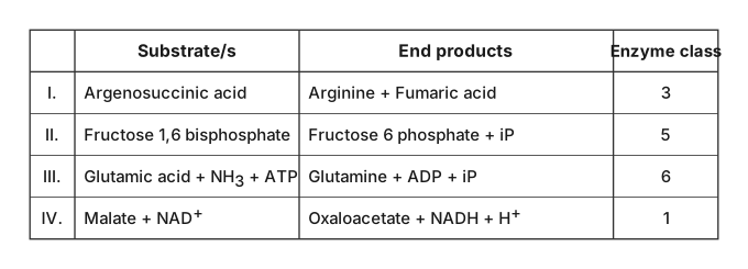
›Reveal solutionSolution
The question tests your ability to match a biochemical reaction with the correct enzyme class (EC number). The correct combination is III and IV, which corresponds to option (B).
The enzyme classification system (EC numbers) groups enzymes by the type of reaction they catalyze, not by the substrate or product alone. The six main classes are:
- Oxidoreductases – transfer electrons (redox reactions)
- Transferases – transfer functional groups
- Hydrolases – cleavage using water
- Lyases – addition/removal of groups without hydrolysis or redox
- Isomerases – geometric or structural rearrangements
- Ligases – joining two molecules using ATP (or similar energy source)
Let’s examine each entry one by one.
-
Entry I: Argenosuccinic acid → Arginine + Fumaric acid
This is a cleavage reaction that does not use water (it’s not a hydrolysis) and does not involve redox. The bond is broken to release fumarate, which is a typical lyase reaction (EC class 4). The table assigns class 3 (hydrolase), which is wrong. So I is incorrect.
-
Entry II: Fructose 1,6-bisphosphate → Fructose 6-phosphate + iP
Here, a phosphate group is removed from the first carbon and transferred to water (inorganic phosphate is released). This is a phosphatase reaction — a subclass of hydrolases (EC class 3). But the table assigns class 5 (isomerase), which is incorrect; the reaction is a dephosphorylation (hydrolysis, class 3), not an isomerization.
Watch outA common mistake is to confuse the removal of a phosphate group with isomerization. Fructose 1,6-bisphosphate to fructose 6-phosphate is not an isomerization — it’s a hydrolysis (phosphatase). So entry II is wrong as given.
Since the class number (5, isomerase) does not match the reaction, entry II is incorrect.
-
Entry III: Glutamic acid + NH₃ + ATP → Glutamine + ADP + iP
This is the synthesis of glutamine from glutamate and ammonia, driven by ATP hydrolysis. Joining two molecules using ATP is the hallmark of a ligase (EC class 6). The table assigns class 6 — correct. So III is correct.
-
Entry IV: Malate + NAD⁺ → Oxaloacetate + NADH + H⁺
This is a dehydrogenation — malate loses two hydrogens (one as hydride to NAD⁺, one as proton). That’s an oxidoreductase reaction (EC class 1). The table assigns class 1 — correct. So IV is correct.
Now, the correct entries are III and IV. But look at the options:
(A) II and III
(B) III and IV
(C) I and IV
(D) II and IV
Option (B) is III and IV. That matches our analysis.
TipAlways check the reaction type, not the substrate name. For example, “argininosuccinate → arginine + fumarate” is a lyase (class 4), not a hydrolase. Memorizing the six classes by their action is faster than memorizing individual reactions.
✓Final answerThe correct combination is III and IV, which corresponds to option (B).
- TG EAPCET 2024Set ap-2024-05-07-FN1 markMCQQ.In which of the following organisms, cell wall is not found in any stage? (A) Dinoflagellates (B) Chrysophytes (C) Slime moulds (D) Protozoans
›Reveal solutionSolution
The key idea is that protozoans are animal-like protists that lack a cell wall entirely, unlike the other groups listed which have cell walls at some life stage. The correct answer is (D).
Concept & Intuition
A cell wall is a rigid outer layer found in plants, fungi, and many protists, providing structural support and protection. However, protozoans are unicellular, heterotrophic organisms that behave like tiny animals—they move, engulf food, and lack photosynthesis. Since animals never have cell walls, protozoans (being animal-like) also lack them in all life stages. The other options—dinoflagellates, chrysophytes, and slime moulds—are all protists that possess cell walls at least during part of their life cycle.
Step-by-step reasoning
-
Dinoflagellates (Option A)
These are mostly marine, photosynthetic protists. They have a distinctive cell wall made of cellulose plates (theca) that forms a rigid armor. This wall is present in both motile and non-motile stages.
→ Cell wall is present.
-
Chrysophytes (Option B)
This group includes diatoms and golden algae. Diatoms have a unique silica-based cell wall (frustule) that is always present. Golden algae may have cellulose or pectin walls.
→ Cell wall is present.
-
Slime moulds (Option C)
Slime moulds are fungus-like protists. During their feeding (plasmodial) stage, they lack a cell wall, but when they form fruiting bodies (sporangia) for reproduction, the spores are encased in a cellulose wall. Thus, a cell wall appears at least in the spore stage.
→ Cell wall is present in some stage.
-
Protozoans (Option D)
Protozoans (e.g., Amoeba, Paramecium, Plasmodium) are heterotrophic, motile protists. They have a flexible cell membrane (pellicle in some) but never a rigid cell wall. This is because they rely on phagocytosis (engulfing food) and movement via pseudopodia, cilia, or flagella—a cell wall would hinder these processes.
→ Cell wall is absent in all stages.
Watch outA common mistake is to think slime moulds have no cell wall because their feeding stage is wall-less. But remember: the spore stage does have a cellulose wall, so they are not “wall-free in any stage.”
TipA quick memory aid: “Protozoans are animal-like → no cell wall; others are plant-like or fungus-like → have cell walls.”
✓Final answerThe correct option is (D).
ANSWER: D
-
- TG EAPCET 2023Set ap-2023-05-10-FN1 markMCQQ.Statins are produced by Monascus purpureus and it is a (A) Algae (B) Bacteria (C) Virus (D) Yeast
›Reveal solutionSolution
Statins are cholesterol-lowering drugs produced by the fungus Monascus purpureus, which is a yeast — so the correct answer is (D).
The question tests your knowledge of a specific microorganism used in industrial biotechnology. Monascus purpureus is a well-known fungus, often called red yeast rice, and it naturally produces statins like lovastatin. Statins work by inhibiting the enzyme HMG-CoA reductase, which is key in cholesterol synthesis.
Let’s break down why each option is right or wrong.
-
Identify the organism. Monascus purpureus is a filamentous fungus, but in common classification it falls under yeasts (ascomycete fungi). It is not an alga, bacterium, or virus. Yeasts are single-celled fungi, and Monascus is a classic example used in fermentation (e.g., red yeast rice in Asian cuisine).
-
Why not algae? Algae are photosynthetic eukaryotes (like Chlorella or Spirogyra). Monascus is non-photosynthetic and heterotrophic — it feeds on organic matter. So (A) is wrong.
-
Why not bacteria? Bacteria are prokaryotes (no nucleus). Monascus has a true nucleus and membrane-bound organelles, making it a eukaryote. So (B) is wrong.
-
Why not virus? Viruses are acellular and require a host to replicate. Monascus is a living, cellular organism that can be cultured on its own. So (C) is wrong.
-
Confirm the yeast connection. In many textbooks, Monascus purpureus is explicitly listed under yeasts (or molds, but molds are also fungi). Statins are produced by this fungus, and the question’s options include “Yeast” as the only fungal category. Therefore, (D) is correct.
Watch outA common mistake is to confuse Monascus purpureus with a bacterium because it’s used in fermentation. Remember: it’s a fungus (yeast), not a prokaryote.
TipIf you ever forget, recall that statins are fungal secondary metabolites — and the only fungal option here is yeast. Algae, bacteria, and viruses don’t produce statins naturally.
✓Final answerThe correct option is (D) Yeast.
-
- TG EAPCET 2023Set ap-2023-05-10-AN1 markMCQQ.Pusa komal for bacterial blight and pusa swarnim for white rust are processed in these crops respectively (A) Wheat and Brassica (B) Cauliflower and chilli (C) Brassica and chilli (D) Cow pea and Brassica
›Reveal solutionSolution
Pusa Komal is a variety of Cowpea resistant to bacterial blight, and Pusa Swarnim is a variety of Brassica resistant to white rust. The correct option is (D).
Plant breeding plays a crucial role in developing crop varieties that are resistant to various diseases, thereby increasing agricultural productivity and reducing reliance on chemical pesticides. This question tests your knowledge of specific improved crop varieties and their associated disease resistances, which are outcomes of such breeding efforts.
Here's how we identify the correct crops:
-
Identify the crop for Pusa Komal:
Pusa Komal is a well-known variety developed through conventional breeding. It is specifically bred for resistance to bacterial blight. This variety belongs to Cowpea (Vigna unguiculata).
-
Identify the crop for Pusa Swarnim:
Pusa Swarnim (also known as Pusa Swarn) is another important variety. It is bred for resistance to white rust, a fungal disease. This variety belongs to Brassica (mustard).
-
Match the crops with the given options:
The question asks for the crops "respectively" for Pusa Komal (bacterial blight) and Pusa Swarnim (white rust).
- Pusa Komal → Cowpea
- Pusa Swarnim → Brassica
Comparing this with the given options:
- (A) Wheat and Brassica - Incorrect, Pusa Komal is not Wheat.
- (B) Cauliflower and chilli - Incorrect.
- (C) Brassica and chilli - Incorrect, Pusa Komal is not Brassica.
- (D) Cow pea and Brassica - Correct, as Pusa Komal is Cowpea and Pusa Swarnim is Brassica.
ImportantKnowing specific examples of disease-resistant varieties and the crops they belong to is essential for understanding the applications of plant breeding.
✓Final answerThe crops for Pusa Komal (bacterial blight) and Pusa Swarnim (white rust) are Cow pea and Brassica respectively, making the correct option (D).
-
- TG EAPCET 2023Set ap-2023-05-11-AN1 markMCQQ.In a rDNA cloning experiment, which of the following helps to identify and eliminate non-transformant normal Escherichia coli cells (A) Origin of Replication (Ori) (B) Selectable marker (C) Cloning site (D) Competent host
›Reveal solutionSolution
In rDNA cloning, selectable markers are genes on a plasmid that allow transformed cells to survive or exhibit a distinct phenotype under specific conditions, thereby identifying and eliminating E. coli cells that did not take up the plasmid (non-transformants). The correct option is (B).
In recombinant DNA (rDNA) cloning, the goal is to introduce a desired gene into a host cell, typically Escherichia coli, using a plasmid vector. However, not all E. coli cells will successfully take up the plasmid during the transformation process. Cells that do not take up the plasmid are called non-transformants. It is crucial to distinguish these non-transformant cells from the transformant cells (those that have taken up the plasmid) and eliminate them, so that only the desired cells are propagated. This is where selectable markers play a vital role.
Here's a breakdown of how this works and why selectable markers are the key:
-
Understanding Transformation and Non-Transformants:
When E. coli cells are treated to become "competent" (capable of taking up foreign DNA), they are mixed with the recombinant plasmid. However, the efficiency of this process is very low; only a small fraction of cells actually take up the plasmid. The cells that do not take up any plasmid are the "non-transformant normal Escherichia coli cells" mentioned in the question. These cells are essentially unchanged and do not contain the desired genetic material.
-
The Need for Selection:
If we were to grow all the cells (transformants and non-transformants) together, the non-transformants, being the vast majority, would quickly outgrow the transformants. This would make it impossible to isolate and propagate the cells containing the recombinant DNA. Therefore, a mechanism is needed to selectively allow only the transformants to grow, while eliminating the non-transformants.
-
Role of Selectable Markers:
A selectable marker is a gene carried on the plasmid vector that confers a specific phenotype (like antibiotic resistance) to the host cell that has taken up the plasmid. When these cells are grown under selective conditions (e.g., in a medium containing the antibiotic), only the cells that possess the plasmid (and thus the selectable marker gene) will survive and multiply. Non-transformant cells, lacking the plasmid and the selectable marker, will be unable to grow and will be eliminated.
Plasmid with selectable marker → Transformant cell → Survives selective conditions
No plasmid (non-transformant) → Dies under selective conditions
A common example is a plasmid carrying an ampicillin resistance gene (ampR). If E. coli cells are transformed with this plasmid and then plated on a medium containing ampicillin, only the cells that have taken up the plasmid will be able to grow, forming colonies. The non-transformant cells, which are sensitive to ampicillin, will die.
-
Evaluating the Options:
-
(A) Origin of Replication (Ori): The Ori is a specific DNA sequence on the plasmid where DNA replication initiates. It ensures that the plasmid can replicate independently within the host cell, maintaining its copy number. While essential for the plasmid's propagation, it does not provide a mechanism to distinguish between transformant and non-transformant cells or to eliminate non-transformants.
-
(B) Selectable marker: As explained above, selectable markers (like genes for antibiotic resistance, e.g., ampicillin resistance, tetracycline resistance) allow for the identification and selection of transformant cells by enabling them to survive under conditions that are lethal to non-transformant cells. This directly helps to identify and eliminate non-transformant normal Escherichia coli cells.
-
(C) Cloning site: Also known as a multiple cloning site (MCS) or polylinker, this is a region on the plasmid containing several recognition sites for different restriction enzymes. It is where the foreign DNA (the gene of interest) is inserted into the plasmid. The cloning site is crucial for inserting the desired gene, but it does not directly help in distinguishing transformants from non-transformants. It is involved in distinguishing recombinants (plasmids with insert) from non-recombinants (plasmids without insert), which is a subsequent step after initial transformation selection.
-
(D) Competent host: A competent host refers to a bacterial cell (like E. coli) that has been treated to increase its permeability to DNA, making it capable of taking up foreign DNA. While a competent host is absolutely necessary for the transformation process itself, it is the recipient of the DNA, not the tool for selection. It does not help in identifying or eliminating non-transformants after the transformation attempt.
-
Therefore, the selectable marker is the component that specifically helps to identify and eliminate non-transformant E. coli cells.
✓Final answerThe selectable marker helps to identify and eliminate non-transformant normal Escherichia coli cells, so the correct option is (B).
-
- TG EAPCET 2023Set ap-2023-05-11-AN1 markMCQQ.Identify A and B in the following equation Casein A,Ca++ Calcium paracaseinate B Peptones (A) A. Renin B. Chymotrypsin (B) A. Rennin B. Trypsin (C) A. Ptyalin B. Pepsin (D) A. Rennin B. Pepsin
›Reveal solutionSolution
The conversion of casein to calcium paracaseinate requires rennin (also called chymosin), and the further breakdown to peptones is done by pepsin. The correct option is (D).
This question tests your knowledge of milk digestion — a specific biochemical pathway that is often confused with general protein digestion. Casein is the main protein in milk, and it has a unique clotting step before it can be fully digested.
The key idea: casein in milk is soluble, but it must first be coagulated (clotted) by the enzyme rennin in the presence of calcium ions. This forms calcium paracaseinate, an insoluble curd. Then, the curd is attacked by pepsin (and later trypsin) to break it into smaller peptides and peptones.
Let’s walk through the steps.
-
First step: Casein → Calcium paracaseinate
The enzyme that clots milk is rennin (also called chymosin). It is secreted by the gastric glands of infants and young mammals. Rennin acts on caseinogen (the soluble form) to form paracasein, which then reacts with Ca++ ions to form insoluble calcium paracaseinate — the curd.
Watch outDo not confuse rennin with renin. Renin is a kidney enzyme involved in blood pressure regulation, not milk digestion. The spelling matters in exams.
-
Second step: Calcium paracaseinate → Peptones
Once the milk is clotted, the curd is further digested by pepsin, an enzyme secreted by the stomach. Pepsin breaks down proteins into smaller fragments called peptones and proteoses. (Later, in the small intestine, trypsin and chymotrypsin continue the digestion.)
-
Eliminating the wrong options
- (A) says Renin (wrong spelling and wrong enzyme) and Chymotrypsin (acts in the small intestine, not on the curd directly).
- (B) says Rennin (correct for first step) but Trypsin (trypsin acts in the small intestine, not in the stomach; it is not the primary enzyme for curd → peptones).
- (C) says Ptyalin (salivary amylase, digests starch, not protein) and Pepsin (correct for second step, but first step is wrong).
- (D) says Rennin and Pepsin — both correct for their respective steps.
TipRemember the sequence: Rennin clots milk → Pepsin digests the clot → Trypsin continues in the intestine. In exams, the first two steps (stomach) are the most frequently tested.
✓Final answerThe correct option is (D): A is Rennin, B is Pepsin.
-
- TG EAPCET 2022Set ap-2022-07-30-FN1 markMCQQ.Choose the correct statement from the following: (A) Gene therapy involves ELISA technique to treat non-functional gene. (B) A and B chains of human insulin was produced separately in E. coli; these were extracted and combined by disulphide bond to form human insulin. (C) Parentage dispute will be solved by RNA interference technique. (D) Transgenic papaya is resistant to Pseudomonas pathogen.
›Reveal solutionSolution
The question tests knowledge of biotechnology applications; only statement (B) correctly describes the production of human insulin in E. coli by separately synthesizing A and B chains and joining them via disulfide bonds.
The key here is to recall specific, well‑established facts from biotechnology — not to guess. Each option refers to a distinct technique or application, and only one matches the actual scientific procedure.
-
Option (A): Gene therapy involves ELISA technique to treat non-functional gene.
- Gene therapy aims to correct a defective gene by introducing a functional copy into the patient’s cells.
- ELISA (Enzyme‑Linked Immunosorbent Assay) is a diagnostic tool for detecting proteins or antibodies — it is not used to deliver or repair genes.
- This statement confuses a detection method with a therapeutic method. Incorrect.
-
Option (B): A and B chains of human insulin was produced separately in E. coli; these were extracted and combined by disulphide bond to form human insulin.
- This is the classic method for producing recombinant human insulin (Humulin).
- The A and B polypeptide chains are synthesized in separate E. coli cultures, then purified and mixed under controlled conditions to form disulfide bridges, yielding active insulin.
- This is a landmark achievement in biotechnology. Correct.
-
Option (C): Parentage dispute will be solved by RNA interference technique.
- RNA interference (RNAi) silences specific genes by degrading mRNA — it is used in research and therapy, not for genetic fingerprinting.
- Parentage disputes are resolved by DNA profiling (e.g., using STR markers), not by RNAi. Incorrect.
-
Option (D): Transgenic papaya is resistant to Pseudomonas pathogen.
- The famous transgenic papaya (Rainbow papaya) is engineered for resistance to Papaya ringspot virus (PRSV), not a bacterial pathogen like Pseudomonas.
- Pseudomonas is a bacterial genus; the resistance in transgenic papaya is viral, not bacterial. Incorrect.
Watch outA common mistake is to confuse “RNA interference” with “DNA fingerprinting” because both involve nucleic acids — but they serve completely different purposes.
TipRemember the insulin story: separate chains in E. coli, then combine in vitro. This is a classic example of post‑translational modification outside a living cell.
✓Final answerThe correct option is (B).
ANSWER: B
-
- TG EAPCET 2022Set ap-2022-07-30-FN1 markMCQQ.Which of the following contains the enzyme lysozyme? (A) Gastric juice (B) Bile juice (C) Saliva (D) Pancreatic juice
›Reveal solutionSolution
Lysozyme is an antibacterial enzyme found in saliva (and also in tears, mucus, and breast milk). The correct option is (C).
The question tests your knowledge of the composition of digestive juices — specifically, which one contains an enzyme that is not primarily for digestion but for defence. Lysozyme breaks down the cell walls of certain bacteria, acting as a first line of protection. Among the options, only saliva is secreted in the mouth, where it mixes with food and also coats the oral cavity, providing both lubrication and antimicrobial action.
Let’s go through each option:
-
Gastric juice — Secreted by the stomach lining. It contains hydrochloric acid (HCl), pepsinogen (activated to pepsin), and mucus. Its main job is to digest proteins and kill microbes with strong acidity. Lysozyme is not a component here.
-
Bile juice — Produced by the liver and stored in the gallbladder. It contains bile salts, bile pigments (like bilirubin), cholesterol, and phospholipids. Bile has no enzymes at all — it emulsifies fats for digestion but does not contain lysozyme.
-
Saliva — Secreted by the salivary glands (parotid, submandibular, sublingual). It contains water, electrolytes, mucus, the digestive enzyme amylase (starts starch breakdown), and lysozyme. Lysozyme attacks peptidoglycan in bacterial cell walls, making saliva a mild antiseptic. This is the correct answer.
-
Pancreatic juice — Released by the pancreas into the small intestine. It contains many digestive enzymes: trypsinogen, chymotrypsinogen, pancreatic amylase, lipase, and nucleases. It also has bicarbonate to neutralise stomach acid. Lysozyme is not present.
Watch outA common mistake is to think that gastric juice contains lysozyme because it kills bacteria. But the stomach uses HCl (strong acid) for that purpose, not lysozyme. Lysozyme is found in secretions that are not strongly acidic, like saliva and tears.
✓Final answerThe correct option is (C) Saliva.
-
- TG EAPCET 2022Set ap-2022-07-30-FN1 markMCQQ.In DNA finger printing technology, the DNA is fragmented by the enzyme (A) Restriction endonuclease (B) Reverse transcriptase (C) DNase (D) RNase
›Reveal solutionSolution
DNA fingerprinting begins by cutting DNA into fragments using restriction endonucleases, which recognize specific sequences and make precise cuts — the correct option is (A).
The entire process of DNA fingerprinting hinges on generating a unique pattern of DNA fragments from an individual’s genome. To get those fragments, you need an enzyme that cuts DNA at specific, reproducible sites. That’s the job of restriction endonucleases — they are the molecular scissors that recognize short, palindromic sequences (like GAATTC) and cut the DNA at those exact points. This produces fragments of varying lengths, which are then separated by gel electrophoresis and probed for repetitive sequences (VNTRs) to create a unique fingerprint.
Let’s walk through why each option stands or falls.
-
Restriction endonuclease — These enzymes are naturally produced by bacteria to defend against viruses. They cut DNA only at specific recognition sites, typically 4–8 base pairs long. In DNA fingerprinting, you need a reproducible, sequence-specific cut to generate consistent fragment patterns. That’s exactly what restriction endonucleases provide. Without them, you cannot get the characteristic banding pattern that distinguishes one individual from another.
-
Reverse transcriptase — This enzyme makes DNA from an RNA template. It’s used in molecular biology to create complementary DNA (cDNA) from mRNA, but it has no role in cutting genomic DNA. In DNA fingerprinting, you start with genomic DNA, not RNA, so reverse transcriptase is irrelevant here.
-
DNase — This is a general term for enzymes that degrade DNA. DNase I, for example, cuts DNA non-specifically, producing a random smear of fragments. That’s the opposite of what you want — DNA fingerprinting requires precise, reproducible cuts at defined sequences. Random degradation would destroy the pattern, not create it.
-
RNase — This enzyme degrades RNA, not DNA. In DNA fingerprinting, you might use RNase to clean up RNA contamination from a sample, but it does not fragment the DNA itself. So it’s not the answer.
Watch outA common mistake is to confuse DNase (non-specific DNA cutter) with restriction endonuclease (sequence-specific cutter). Remember: DNase gives a random mess; restriction endonuclease gives a reproducible pattern — only the latter is useful for fingerprinting.
TipIn the lab, you’ll often see the enzyme written as “restriction endonuclease” or simply “restriction enzyme.” The key exam point: it cuts DNA at specific palindromic sequences, producing sticky or blunt ends. That specificity is what makes DNA fingerprinting possible.
✓Final answerThe correct option is (A) Restriction endonuclease.
-
🎓Unlock everything free for 14 days
- ✓Full step-by-step solutions
- ✓Concept-first explanations
- ✓Methods, shortcuts & mistakes
- ✓PYQ mapping + timed mock tests
Full access for 14 days. No credit card required.