Q.Do you know for how many months pregnancy last in dogs, elephants, cats? Find out.
🔒You're viewing a preview — the full solution, concept, methods & PYQ mapping are locked.
🔒 Start your 14-day free trial to unlock the full solution →Concept understanding — Human Reproduction
Human Reproduction: A First Look
Think about what makes you you. You have your mother's eyes, your father's laugh, a mix of traits from two people you may never have met — your grandparents. That chain of life, stretching back thousands of years, runs through one process: human reproduction. It is not a topic for biology students alone. It is the story of how every single person on this planet came to be here.
The Big Picture: Why Does Reproduction Exist?
Every living thing has one fundamental drive: to continue its kind. A mango tree grows fruit so its seeds can sprout elsewhere. A bacterium splits in two. For humans, reproduction is more complex — and more personal. It is the reason families exist, why you have siblings or cousins, and why populations grow or shrink.
At its simplest, human reproduction is the biological process by which a new human individual is created. It requires two parents — a male and a female — each contributing a special cell that fuses to form a single, new cell. That single cell then grows, divides, and develops into a baby over about nine months.
Reproduction is not the same as sex. Sex is an act; reproduction is the entire sequence from cell fusion to birth. Not every act of sex leads to reproduction, and reproduction can sometimes happen with medical help (like IVF) without sex.
The Two Key Players: Male and Female Reproductive Systems
Your body, whether male or female, has a set of organs designed specifically for reproduction. These are not the same as the organs that keep you alive — they are the "spare set" dedicated to creating the next generation.
The Male System: Production and Delivery
The male reproductive system has one main job: to produce male sex cells (sperm) and deliver them to the female body. The key parts are:
- Testes (two oval organs in a pouch called the scrotum): These are the factories. They produce sperm continuously from puberty onwards — millions every day. They also produce testosterone, the hormone that drives male physical changes at puberty.
- Duct system (a series of tubes): Sperm travel from the testes through a long coiled tube (epididymis) where they mature, then through a muscular tube (vas deferens) that carries them toward the urethra.
- Accessory glands (seminal vesicles, prostate gland, bulbourethral glands): These add fluids to the sperm to nourish them and help them swim. The mixture of sperm and these fluids is called semen.
- Penis: The organ that delivers semen into the female reproductive tract.
The Female System: Production, Reception, and Nurturing
The female system has a bigger job: it produces female sex cells (eggs), receives sperm, and if fertilization occurs, houses and nourishes the developing baby for nine months. The key parts are:
- Ovaries (two almond-sized organs): These produce eggs — but unlike sperm, eggs are not made continuously. A female is born with all the eggs she will ever have (about 1–2 million at birth, reducing to around 300,000–400,000 by puberty). Each month, one egg matures and is released.
- Fallopian tubes (also called oviducts): These are not connected directly to the ovaries. When an egg is released, the finger-like ends of the tube sweep it inside. Fertilization — the meeting of sperm and egg — happens here, in the tube.
- Uterus (womb): A hollow, pear-shaped muscular organ. If fertilization happens, the embryo implants in the lining of the uterus and grows here. If not, the lining is shed — that is menstruation.
- Cervix: The lower, narrow part of the uterus that opens into the vagina. It produces mucus that changes consistency during the month to help or hinder sperm.
- Vagina: A muscular canal that receives the penis during intercourse and serves as the birth canal during delivery.
The female reproductive system has a monthly cycle (the menstrual cycle) that prepares the body for a possible pregnancy. This cycle is controlled by hormones and typically lasts about 28 days. The release of the egg (ovulation) happens around day 14. If no pregnancy occurs, the uterine lining is shed as menstrual blood — this is a normal, healthy process, not a "curse" or something to be ashamed of.
The Moment of Creation: Fertilization
When sperm from the male meet the egg from the female inside a fallopian tube, one sperm may penetrate the egg's outer layer. That single moment — fertilization — creates a new cell called a zygote. This zygote contains 23 chromosomes from the mother and 23 from the father, making 46 in total. That is the complete genetic blueprint for a new human being.
The zygote immediately begins to divide as it travels down the fallopian tube toward the uterus. By the time it reaches the uterus (about 5–7 days later), it has become a ball of about 100 cells called a blastocyst. This blastocyst burrows into the soft, thick lining of the uterus — a process called implantation. That is when pregnancy truly begins.
The Nine-Month Journey: Development
Once implanted, the developing human is called an embryo for the first eight weeks, and then a fetus from the ninth week until birth. During these months:
- A special organ called the placenta develops, connecting the mother's blood supply to the fetus. It delivers oxygen and nutrients and removes waste — without the mother's and baby's blood ever mixing directly.
- The umbilical cord links the baby to the placenta.
- The baby is protected by a fluid-filled sac (amniotic sac) that cushions it.
- All major organs form in the first three months (first trimester). The next three months (second trimester) are about growth and refinement. The final three months (third trimester) are about rapid weight gain and preparation for life outside the womb. …
The NCERT textbook does not answer this itself — it is a "find out" prompt for outside research, not content the exam tests. For reference, typical gestation periods are: dog — about 2 months (~63 days); cat — about 2 months (~63-65 days); elephant — about 18-22 months (~620-660 days), the longest gestation period of any land mammal. …
This is an open "find out for yourself" prompt — NCERT does not state the answer. Real-world gestation periods: dog and cat are both around 2 months, while the elephant's gestation (~18-22 months) is the longest of any land mammal.
This question is explicitly framed by the textbook as something for the student to research independently ("Find out"), immediately after noting that human pregnancy lasts about 9 months — it is meant to prompt a comparison, not to test a memorised NCERT fact, so nothing here is graded exam content.
For reference, the approximate real gestation periods (general zoological knowledge, outside the NCERT text) are:
- Dog: about 58-68 days (roughly 2 months).
- Cat: about 63-65 days (roughly 2 months) — similar to the dog. …
Showing the 12 most recent of 44 on this concept.
- TG EAPCET 2026Set ap-2026-05-04-AN1 markMCQQ.Match the followingThe correct answer is (A) A – V, B – III, C – IV, D – II (B) A – II, B – IV, C – I, D – V (C) A – III, B – I, C – IV, D – V (D) A – V, B – I, C – IV, D – II
Type of circulation Examples A Open type I Pisces B Single circulation II Mammals C Incomplete double circulation III Parazoans D Double circulation IV Amphibians V Echinoderms 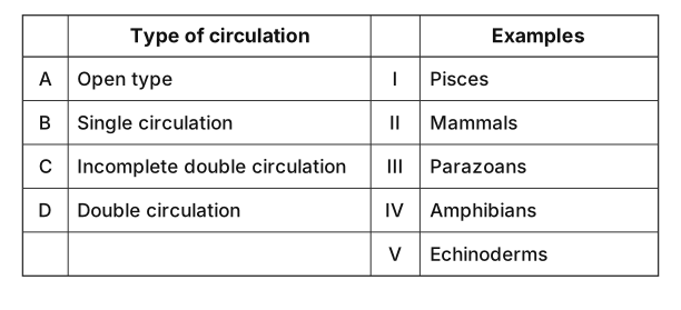
›Reveal solutionSolution
Open type → Echinoderms (V), Single circulation → Pisces (I), Incomplete double → Amphibians (IV), Double → Mammals (II). That is A–V, B–I, C–IV, D–II, i.e. option (D).
The concept first: two independent questions about any circulatory system
Question 1 — is the blood always inside vessels?
- Open circulation: the heart pumps blood into vessels that soon open into large body spaces (sinuses / haemocoel), where the tissues are bathed directly; the fluid then trickles back to the heart. Pressure is low and flow is slow. Found in arthropods (cockroach, prawn), most molluscs, and echinoderms — in a starfish the haemal system is open, and the associated water-vascular system likewise floods open channels.
- Closed circulation: blood is confined to vessels throughout (arteries → capillaries → veins). Pressure is higher and distribution can be regulated precisely. Found in annelids, cephalopods and all vertebrates.
Question 2 — how many times does the blood pass through the heart in one complete circuit?
- Single circulation — once. In a fish, the two-chambered heart (one atrium + one ventricle) receives only deoxygenated blood, pumps it to the gills to be oxygenated, and the oxygenated blood then goes straight on to the body without returning to the heart first. So: heart → gills → body → heart. One pass.
- Double circulation — twice: a pulmonary circuit (heart → lungs → heart) and a systemic circuit (heart → body → heart). It comes in two grades:
- Incomplete double circulation — the heart has three chambers (two atria, one ventricle). Oxygenated blood from the lungs and deoxygenated blood from the body both enter that single ventricle and mix. This is the condition in amphibians (frog) and most reptiles.
- Complete double circulation — the heart has four chambers (two atria, two ventricles), so the two blood streams are completely separated and never mix, giving highly efficient oxygen delivery. This is the condition in birds and mammals. …
- TG EAPCET 2026Set ap-2026-05-04-FN1 markMCQQ.Characters related to ovule in Helianthes I. Inverted II. Curvature angle is 180∘ III. Unitegmic IV. Micropyle lies close to funicle (A) I and II only (B) II and III only (C) I, II, III and IV (D) I, III and IV only
›Reveal solutionSolution
The ovule of Helianthus (sunflower) is anatropous — it is inverted, with a 180∘ curvature, unitegmic, and its micropyle lies close to the funicle. All four statements are correct, so the answer is (C).
The question tests your knowledge of ovule types in angiosperms, specifically in Helianthus (sunflower), a member of the Asteraceae family. In botany, ovules are classified by their orientation and structure. The most common type in flowering plants — and the one found in Helianthus — is the anatropous ovule. Understanding what "anatropous" means is the key to evaluating each statement.
An anatropous ovule is one that has undergone a complete 180° inversion during development. This inversion brings the micropyle (the opening through which the pollen tube enters) close to the funicle (the stalk attaching the ovule to the ovary wall). Such ovules are typically unitegmic (having a single integument) in many advanced families like Asteraceae.
Let’s check each statement:
-
Inverted — In an anatropous ovule, the body of the ovule is turned completely upside down relative to the funicle. The nucellus and embryo sac are oriented such that the micropyle points downward toward the placenta. This is true for Helianthus.
-
Curvature angle is 180∘ — The "curvature angle" refers to the angle between the funicle and the body of the ovule. In an anatropous ovule, this angle is exactly 180∘, meaning the ovule is fully reflexed. This is a defining feature. …
-
- TG EAPCET 2026Set ap-2026-05-04-FN1 markMCQQ.Choose the mismatched pair regarding cockroach (A) Spiracles – taenidia (B) Stigmata – peritremes (C) Trachea – intima (D) Ootheca – collaterial glands
›Reveal solutionSolution
The question tests your knowledge of cockroach anatomy — specifically which structure is not correctly paired with its associated part. The mismatched pair is (D) Ootheca – collaterial glands, because the ootheca is formed by the colleterial glands, not the collaterial glands (a misspelling of the same term). All other pairs are correct.
The key here is to recall the respiratory and reproductive systems of the cockroach. Each option pairs a structure with a feature or associated part. Let's go through them one by one.
-
Spiracles – taenidia: Spiracles are the external openings of the tracheal system. Taenidia are the spiral thickenings inside the tracheae that prevent them from collapsing. This pairing is correct — taenidia are indeed found in the tracheae, which connect to spiracles.
-
Stigmata – peritremes: Stigmata is another name for spiracles. Peritremes are the ring-like sclerites surrounding each spiracle. This is a correct match — peritremes are the structural borders of the stigmata.
-
Trachea – intima: The trachea is lined by a cuticular layer called the intima, which is shed during moulting. This is correct — the intima is the inner lining of the tracheal tubes.
-
Ootheca – collaterial glands: The ootheca is the egg case produced by the female cockroach. It is secreted by the colleterial glands (accessory reproductive glands). The term "collaterial glands" is a common misspelling or variant of "colleterial glands". However, in standard biology, the correct spelling is colleterial glands. Since the option uses "collaterial" (which is not the standard term), and more importantly, the pairing is actually correct in function — the ootheca is formed by the colleterial glands. But wait: the question asks for the mismatched pair. Let's re-examine carefully.
Watch outA common trap: students often think "collaterial" is a typo and mark this as mismatched. But in many textbooks, "collaterial glands" is used interchangeably with "colleterial glands". The real mismatch here is subtle — the ootheca is indeed formed by these glands, so the pair is actually correct. That means the mismatched pair must be one of the others. …
-
- TG EAPCET 2026Set ap-2026-05-04-FN1 markMCQQ.Study the following and identify the correct statements I. During inspiration thoracic spiracles are kept closed in cockroach II. In cockroach oenocytes of fat bodies contain symbiotic bacteria III. In cockroach stomodael valve lies between gizzard and mesenteron IV. In cockroach, alary muscles keep the blood in circulation (A) I, II (B) III, IV (C) I, IV (D) II, III
›Reveal solutionSolution
The question tests four statements about cockroach anatomy and physiology. The correct statements are III (stomodael valve lies between gizzard and mesenteron) and IV (alary muscles keep blood in circulation), so the answer is option (B).
The cockroach is a classic example in insect morphology, and these statements touch on respiration, fat body function, digestive tract structure, and circulation. Let’s examine each one carefully, because exam questions often mix a plausible-sounding wrong statement with a correct one.
-
Statement I: “During inspiration thoracic spiracles are kept closed in cockroach.”
Cockroaches have a tracheal system for gas exchange. Spiracles (openings on the thorax and abdomen) are controlled by valves. During inspiration (air intake), the abdominal spiracles typically open while the thoracic spiracles close, and during expiration the reverse happens. This is a coordinated mechanism to create a unidirectional airflow. So the statement is correct — thoracic spiracles are indeed kept closed during inspiration.
But wait — many textbooks state that in cockroaches, the first pair of thoracic spiracles (on the prothorax) are always open, while the others are valved. However, the standard NCERT description says: during inspiration, the abdominal spiracles open and thoracic spiracles close. So this statement is true as per the syllabus.
-
Statement II: “In cockroach oenocytes of fat bodies contain symbiotic bacteria.”
Oenocytes are cells associated with the fat body in insects, and they are involved in lipid metabolism and cuticle formation. They do not contain symbiotic bacteria. Symbiotic bacteria in cockroaches are found in specialized cells called mycetocytes (or bacteriocytes) within the fat body, not in oenocytes. This is a common confusion — oenocytes and mycetocytes are different cell types. So statement II is false.
-
Statement III: “In cockroach stomodael valve lies between gizzard and mesenteron.”
The cockroach digestive tract has three regions: foregut (stomodeum), midgut (mesenteron), and hindgut (proctodeum). The stomodael valve (also called the cardiac valve or proventricular valve) is indeed located at the junction of the gizzard (part of foregut) and the mesenteron (midgut). It regulates the passage of food. This is a standard fact — statement III is correct.
-
Statement IV: “In cockroach, alary muscles keep the blood in circulation.” …
-
- TG EAPCET 2026Set ap-2026-05-04-FN1 markMCQQ.Consider the following statements Statement I: Both anal and sphincters are formed by smooth muscles in man Statement II: Renin converts the milk into curd (A) Both statement I and statement II are true (B) Both statement I and statement II are false (C) Statement I is true, but statement II is false (D) Statement I is false, but statement II is true
›Reveal solutionSolution
Statement I is false because the external anal sphincter is made of skeletal muscle, not smooth muscle. Statement II is false because renin is an enzyme involved in blood pressure regulation, not milk curdling. Both statements are false.
The key to this question is knowing the precise composition of the anal sphincters and the specific function of renin. Many students confuse the two anal sphincters (internal vs. external) and also mix up renin with rennin (also called chymosin), which is the enzyme that actually curdles milk. Let’s break each statement down.
-
Statement I: "Both anal sphincters are formed by smooth muscles in man"
The anal canal has two sphincters: the internal anal sphincter and the external anal sphincter. The internal anal sphincter is made of smooth muscle (involuntary, under autonomic control). The external anal sphincter, however, is made of skeletal muscle (voluntary, under somatic control). Because the external sphincter is not smooth muscle, the statement that both are formed by smooth muscle is false.
-
Statement II: "Renin converts the milk into curd" …
-
- TG EAPCET 2026Set ap-2026-05-04-FN1 markMCQQ.Consider the following statements Assertion (A): Placenta of human beings is described as haemochorial type Reason (R): Chorionic villi of the foetus are dipped in the blood pools of uterine wall (A) Both (A) and (R) are correct, (R) is the correct explanation for (A) (B) Both (A) and (R) are correct, (R) is not the correct explanation for (A) (C) (A) is correct, but (R) is not correct (D) (A) is not correct, but (R) is correct
›Reveal solutionSolution
The human placenta is haemochorial because the foetal chorionic villi are directly bathed in maternal blood pools. Both statements are correct, and the Reason correctly explains the Assertion.
The question tests your understanding of placental classification in humans. The term "haemochorial" literally means "blood" (haemo-) and "chorion" (the foetal membrane). The key idea is that in this type of placenta, the maternal blood comes into direct contact with the foetal chorionic tissue — there is no intervening layer of maternal endothelium or uterine epithelium. The Reason describes exactly that: the chorionic villi (foetal tissue) are dipped into pools of maternal blood. So the Reason is not just a true fact; it is the very definition of why we call it haemochorial.
Let’s walk through the logic step by step.
-
What does "haemochorial" mean?
In placental mammals, the placenta is classified by how many layers separate maternal and foetal blood. In the haemochorial type, the maternal blood directly bathes the chorionic villi. The foetal chorion is in direct contact with maternal blood — hence "haemo" (blood) + "chorion". Humans, along with rodents and some other mammals, have this type.
-
Check the Assertion (A):
"Placenta of human beings is described as haemochorial type."
This is correct. In human pregnancy, the trophoblast (outer layer of the blastocyst) erodes into the maternal endometrium, creating spaces called lacunae that fill with maternal blood. The chorionic villi project into these blood-filled spaces. So the assertion is true.
-
Check the Reason (R):
"Chorionic villi of the foetus are dipped in the blood pools of uterine wall." …
-
- TG EAPCET 2026Set ap-2026-05-05-FN1 markMCQQ.Zygote is diploid in which life cycle? I. Haplotonic II. Diplontic III. Haplodiplontic IV. Diplohaplontic (A) I, II and III only (B) III and IV only (C) I, II, III and IV (D) I, II and IV only
›Reveal solutionSolution
The zygote is formed by the fusion of two haploid gametes, making it inherently diploid (2n) in all sexually reproducing organisms, regardless of the specific life cycle pattern. Therefore, the zygote is diploid in haplotonic, diplontic, haplodiplontic, and diplohaplontic life cycles. The correct option is (C).
Concept and Intuition
Sexual reproduction fundamentally involves the fusion of two specialized cells called gametes. These gametes are typically haploid, meaning they contain a single set of chromosomes, denoted as n. When two haploid gametes fuse, their nuclei combine, resulting in a cell with two sets of chromosomes. This newly formed cell is called a zygote, and it is always diploid, denoted as 2n.
The type of life cycle (haplontic, diplontic, or haplodiplontic/alternation of generations) describes the relative prominence of the haploid and diploid phases in an organism's overall life history, and when meiosis occurs. However, the formation of the zygote itself is a consistent event: it is always the product of gamete fusion and is therefore always diploid. The subsequent development of the zygote and the timing of meiosis are what define the different life cycle types, not the ploidy of the zygote itself.
Step-by-Step Explanation
-
Understanding Zygote Formation:
A zygote is the first diploid cell formed when two haploid gametes (sperm and egg in many organisms) fuse during fertilization. This fusion is a universal event in sexual reproduction.
-
Ploidy of Gametes:
Gametes are always haploid (n). This means they contain half the number of chromosomes found in the somatic (body) cells of the parent organism. This reduction in chromosome number occurs through a specialized cell division called meiosis.
-
Determining Ploidy of the Zygote:
When a haploid male gamete (n) fuses with a haploid female gamete (n), their genetic material combines. The resulting cell, the zygote, therefore contains two sets of chromosomes (n+n=2n). This makes the zygote inherently diploid.
Haploid Gamete(n)+Haploid Gamete(n)→Diploid Zygote(2n)
-
Applying to Different Life Cycles:
Let's examine how this applies to the life cycles mentioned:
-
I. Haplotonic Life Cycle:
In this life cycle (e.g., many algae, fungi), the dominant phase is haploid. The haploid organism produces haploid gametes by mitosis. These gametes fuse to form a diploid zygote (2n). Crucially, this zygote immediately undergoes meiosis to produce haploid spores, which then develop into new haploid organisms. Even though the diploid phase is very short-lived, the zygote itself is diploid.
-
II. Diplontic Life Cycle:
In this life cycle (e.g., humans, most animals, some algae), the dominant phase is diploid. The diploid organism produces haploid gametes by meiosis. These gametes fuse to form a diploid zygote (2n), which then develops by mitosis into a new diploid organism. Here, the zygote is clearly diploid and initiates the diploid phase.
-
III. Haplodiplontic / IV. Diplohaplontic Life Cycle (Alternation of Generations): …
-
-
- TG EAPCET 2026Set ap-2026-05-05-FN1 markMCQQ.Choose the tautonyms from the following I. Homo sapiens sapiens II. Axis axis III. Corvus splendens splendens IV. Naja naja (A) I, II only (B) I, II, III, IV (C) I, III, IV only (D) II, IV only
›Reveal solutionSolution
Tautonyms are scientific names where the genus and specific epithet are identical, a practice allowed only in zoological nomenclature; thus, only Axis axis and Naja naja are tautonyms among the given options. The correct option is (D).
The Art of Naming: Understanding Tautonyms
In the fascinating world of biological classification, every known species gets a unique scientific name, a system designed to avoid confusion across languages and regions. This system, primarily developed by Carl Linnaeus, is called binomial nomenclature, meaning each species name has two parts: the genus name and the specific epithet (often informally called the species name). For example, in Homo sapiens, Homo is the genus and sapiens is the specific epithet.
Sometimes, we need to distinguish between different populations within a species, leading to trinomial nomenclature for subspecies. Here, a third name, the subspecific epithet, is added. For instance, Homo sapiens sapiens refers to modern humans, where the third sapiens denotes the subspecies.
Now, let's talk about tautonyms. A tautonym is a special case in zoological nomenclature where the genus name and the specific epithet are identical. It's like saying "Deer Deer" or "Cobra Cobra" in a formal scientific way. This practice is only permitted under the International Code of Zoological Nomenclature (ICZN). Interestingly, the International Code of Nomenclature for algae, fungi, and plants (ICNafp) forbids tautonyms.
The intuition behind why they exist often comes from historical naming conventions, where a common name for an animal was simply duplicated when formalized into a scientific name.
Step-by-Step Analysis
Let's examine each given name to determine if it fits the definition of a tautonym. Remember, we're looking for identical genus and specific epithet.
-
Understanding the Structure of Scientific Names
- A binomial name consists of Genus + specific epithet (e.g., Canis lupus).
- A trinomial name consists of Genus + specific epithet + subspecific epithet (e.g., Canis lupus familiaris).
- A tautonym occurs when the Genus name is identical to the specific epithet (e.g., Gorilla gorilla).
-
Analyzing I. Homo sapiens sapiens
- Genus: Homo
- Specific epithet: sapiens
- Subspecific epithet: sapiens
- Here, the specific epithet (sapiens) is identical to the subspecific epithet (sapiens). However, for a name to be a tautonym, the genus name must be identical to the specific epithet. In this case, Homo is not the same as sapiens.
- Therefore, Homo sapiens sapiens is not a tautonym.
Watch outA common pitfall is to confuse a repeated specific epithet in a trinomial (like sapiens sapiens) with a true tautonym, which requires the genus and specific epithet to be identical.
-
Analyzing II. Axis axis
- Genus: Axis …
-
- TG EAPCET 2026Set ap-2026-05-05-FN1 markMCQQ.Consider the following statements Statement I: Radially symmetrical animals are generally sluggish Statement II: In human body, kidneys are retroperitoneal organs The correct answer is Options : (A) Both statement I and statement II are true (B) Both statements I and statement II are false (C) Statement I is true, but statement II is false (D) Statement I is false, but statement II is true
›Reveal solutionSolution
Radially symmetrical animals are typically slow-moving or sessile, and kidneys in the human body are indeed located behind the peritoneum. Therefore, both statements are true.
The question asks us to evaluate two independent biological statements. We need to determine the truthfulness of each statement based on established biological principles.
Concept and Intuition
For Statement I (Radial Symmetry):
Symmetry in animals refers to the arrangement of body parts around a central axis or plane. Radial symmetry means that the body can be divided into two identical halves by any plane passing through the central axis. This body plan is often associated with animals that are sessile (attached to a substrate) or planktonic (drifting), as it allows them to detect food or threats from any direction. Such animals typically do not exhibit fast, directed movement.
For Statement II (Retroperitoneal Organs):
The peritoneum is a serous membrane that lines the abdominal cavity and covers most of the abdominal organs. Organs can be classified based on their relationship to this membrane:
- Intraperitoneal organs are suspended within the peritoneal cavity by mesenteries and are almost completely covered by visceral peritoneum.
- Retroperitoneal organs are located behind the peritoneum, meaning they are covered by peritoneum only on their anterior (front) surface. They are not suspended by mesenteries.
Let's analyze each statement:
- Analyzing Statement I: Radially symmetrical animals are generally sluggish.
- Animals with radial symmetry, such as jellyfish (Cnidaria) and adult starfish (Echinodermata), typically lack a distinct head and tail, which are features associated with directed, active movement.
- Their body plan allows them to interact with their environment from all sides. This is advantageous for sessile organisms (like sea anemones) that filter feed, or for slow-moving or drifting organisms (like jellyfish) that capture prey as it comes into contact with their tentacles. …
- TG EAPCET 2026Set ap-2026-05-05-FN1 markMCQQ.Choose the mismatched pair regarding cockroach (A) Alary muscles – blood circulation (B) Malpighian tubules - excretion (C) Dorso ventral muscles - respiration (D) Anal styles – female
›Reveal solutionSolution
The question asks to identify the mismatched pair regarding cockroach anatomy and function. Anal styles are characteristic structures found only in male cockroaches, not females. Therefore, the pair "Anal styles – female" is mismatched.
The question tests your knowledge of the specific anatomical structures and their functions in a cockroach, particularly focusing on distinguishing features and physiological roles. To identify the mismatched pair, we need to evaluate each option based on established biological facts about cockroaches.
-
Analyze option (A) Alary muscles – blood circulation:
- Alary muscles are fan-shaped muscles located in the dorsal part of the cockroach's body, associated with the dorsal diaphragm.
- Their rhythmic contractions help in the circulation of hemolymph (insect blood) within the hemocoel by assisting the pumping action of the tubular heart. They create pressure gradients that facilitate the movement of hemolymph.
- This pair is matched.
-
Analyze option (B) Malpighian tubules - excretion:
- Malpighian tubules are the primary excretory and osmoregulatory organs in insects, including cockroaches.
- They are fine, yellow, filamentous tubules that absorb metabolic wastes, salts, and water from the hemolymph and discharge them into the hindgut for elimination.
- This pair is matched.
-
Analyze option (C) Dorso ventral muscles - respiration:
- Dorso-ventral muscles (also known as tergo-sternal muscles) connect the dorsal (tergum) and ventral (sternum) plates of the abdominal segments.
- Their contraction and relaxation cause changes in the volume of the abdomen. These abdominal pumping movements create pressure differences within the tracheal system, which aids in the movement of air (ventilation) into and out of the spiracles and tracheae, thereby facilitating respiration.
- While the primary respiratory structures are spiracles and the tracheal system, these muscles play a role in active ventilation, especially during increased metabolic activity. …
-
- TG EAPCET 2026Set ap-2026-05-05-FN1 markMCQQ.Consider the following statements Statement I: Main region of digestion in stomach in man is pyloric part Statement II: Lipase of bile juice play an important role in digestion of fats The correct answer is (A) Both statement I and statement II are true (B) Both statement I and statement II are false (C) Statement I is true, but statement II is false (D) Statement I is false, but statement II is true
›Reveal solutionSolution
The question tests two separate biology facts: the stomach’s main digestive region and the source of lipase. Statement I is false (the pyloric part is mainly for chyme passage, not digestion); Statement II is false (bile has no lipase; pancreatic lipase does the work). So both are false.
Concept & Intuition
This is a classic “two-statement true/false” question from human physiology. You need to recall two distinct ideas:
- The stomach has different regions (cardiac, fundic, body, pyloric). The main site of chemical digestion in the stomach is the body (or fundus), where gastric glands secrete pepsinogen and HCl. The pyloric part is mostly a muscular funnel that regulates chyme release into the duodenum — it does very little digestion.
- Bile is produced by the liver, stored in the gallbladder, and released into the small intestine. It contains bile salts (for fat emulsification) but no enzymes. Lipase, the fat-digesting enzyme, comes from the pancreas (pancreatic lipase) and, to a lesser extent, from the stomach (gastric lipase). Bile juice itself has no lipase.
Thus both statements are incorrect.
Step-by-step reasoning
-
Evaluate Statement I: “Main region of digestion in stomach in man is pyloric part”
- The stomach is divided into cardia, fundus, body, and pyloric part.
- The body (and fundus) contains chief cells (secrete pepsinogen) and parietal cells (secrete HCl). This is where protein digestion begins.
- The pyloric part (pyloric antrum and pyloric canal) secretes mucus and the hormone gastrin, but its primary role is mechanical — mixing and pushing chyme into the duodenum.
- Therefore, the pyloric part is not the main region of digestion; the body is.
- Conclusion: Statement I is false.
-
Evaluate Statement II: “Lipase of bile juice play an important role in digestion of fats”
- Bile juice contains water, bile salts, bilirubin, cholesterol, and electrolytes — but no digestive enzymes. …
- TG EAPCET 2026Set ap-2026-05-05-FN1 markMCQQ.Consider the following Assertion (A): Colostrum is absolutely essential for the newly born baby Reason (R): It contains several antibodies that protect the new born from initial sources of infections (A) Both (A) and (R) are correct, (R) is the correct explanation of (A) (B) Both (A) and (R) are correct, (R) is not the correct explanation of (A) (C) (A) is correct, but (R) is not correct (D) (A) is not correct, but (R) is correct
›Reveal solutionSolution
Colostrum is essential because it provides passive immunity via maternal antibodies. Both the assertion and reason are true, and the reason correctly explains the assertion — so option (A) is correct.
The question tests your understanding of colostrum and its role in passive immunity — a key concept in human physiology and immunology. Let’s break it down.
-
What is colostrum?
Colostrum is the first milk secreted by the mother immediately after childbirth, typically for the first few days. It is thick, yellowish, and rich in nutrients, but its most critical component is antibodies (immunoglobulins), especially IgA.
-
Why is it essential for a newborn?
A newborn’s immune system is immature and cannot produce its own antibodies effectively. The antibodies in colostrum are absorbed directly into the baby’s bloodstream through the gut, providing passive immunity — immediate protection against pathogens the mother has encountered. This is vital because the baby is suddenly exposed to a world full of microbes after the sterile environment of the womb.
-
Does the reason correctly explain the assertion?
The assertion says colostrum is “absolutely essential” — and it is, because without those maternal antibodies, the newborn would be highly vulnerable to infections. The reason states that colostrum contains antibodies that protect against initial infections. This is the direct mechanism by which colostrum provides that essential protection. So the reason is not just true — it is the specific explanation for why the assertion holds. …
-
🎓Unlock everything free for 14 days
- ✓Full step-by-step solutions
- ✓Concept-first explanations
- ✓Methods, shortcuts & mistakes
- ✓PYQ mapping + timed mock tests
Full access for 14 days. No credit card required.