Question of 27
Q.Draw a labelled diagram of the T.S of the spinal cord of man.
Telangana TsbieTelangana Board of Intermediate Education 2023Subjective· 4mImportance★★★★★
0% · 0/27 Questions
You're viewing a preview — the full solution, concept, methods & PYQ mapping are locked.
Start your 14-day free trial to unlock the full solution →A T.S. of the spinal cord shows central H-shaped grey matter (with a central canal, dorsal and ventral horns) surrounded by white matter, plus the dorsal root with its ganglion, the ventral root, and the spinal nerve; label the median sulcus and fissure and meninges.
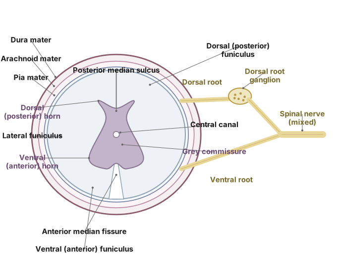
The spinal cord is a cylindrical part of the central nervous system running inside the vertebral column. In transverse section (T.S.), the following parts are seen and should be labelled:
- Central canal: a small canal in the centre, filled with cerebrospinal fluid.
- Grey matter: arranged around the central canal in the shape of the letter H (or a butterfly). It projects as the dorsal (posterior) horns and the ventral (anterior) horns. (Grey matter is made mainly of cell bodies.)
- White matter: surrounds the grey matter; made of myelinated nerve fibres (tracts), it is the outer region.
- Dorsal median sulcus and ventral median fissure: shallow groove on the dorsal side and a deep fissure on the ventral side, partly dividing the cord into two halves.
- Dorsal root and dorsal root ganglion: the dorsal (posterior) root carries sensory fibres and bears a swelling, the dorsal root ganglion.
- Ventral root: the ventral (anterior) root carries motor fibres.
- Spinal nerve: the dorsal and ventral roots join laterally to form a mixed spinal nerve.
- Meninges: the protective membranes (dura mater, arachnoid, pia mater) covering the cord. …
Unlock everything free for 14 days
- Full step-by-step solutions
- Concept-first explanations
- Methods, shortcuts & mistakes
- PYQ mapping + timed mock tests
Full access for 14 days. No credit card required.