Biology · Ch 9 — Biotechnology: Principles and Processes
Cloning Vectors
Cloning Vectors
A cloning vector is a DNA molecule used as a vehicle to carry a foreign DNA fragment into a host cell, where it can be replicated and multiplied. The key insight is that certain natural DNA molecules — plasmids and bacteriophages — can replicate independently inside bacterial cells, without relying on the bacterial chromosome. This ability makes them ideal starting points for building artificial vectors.
Plasmids are small, circular extra-chromosomal DNA molecules found in bacteria. Their copy number per cell varies: some plasmids exist in just one or two copies, while others can have 15–100 copies, and under certain conditions, even more. Bacteriophages (viruses that infect bacteria) have a very high copy number per cell because they produce many viral particles during infection. If you link a foreign piece of DNA to a plasmid or bacteriophage DNA, that foreign DNA will be replicated along with the vector, producing as many copies as the vector itself makes. Modern vectors are engineered to make it easy to insert foreign DNA and to distinguish cells that have taken up the recombinant vector (transformants) from those that have not (non-transformants).
A typical cloning vector has three essential features:
Three essential features of a cloning vector:
- Origin of replication (ori)
- Selectable marker
- Cloning sites (restriction sites)
(i) Origin of replication (ori)
This is a specific DNA sequence where replication begins. Any DNA fragment linked to this sequence can be replicated inside the host cell. The ori also controls the copy number of the linked DNA. If you want to recover many copies of your target DNA, you should clone it into a vector whose ori supports a high copy number.
(ii) Selectable marker
A selectable marker is a gene that helps identify and eliminate non-transformants while selectively allowing the growth of transformants. Transformation is the process by which a piece of DNA is introduced into a host bacterium.
For E. coli, the most useful selectable markers are genes that confer resistance to antibiotics such as ampicillin, chloramphenicol, tetracycline, or kanamycin. Normal E. coli cells do not carry resistance to any of these antibiotics, so only cells that have taken up the vector (and therefore the resistance gene) can survive on a medium containing that antibiotic.
(iii) Cloning sites
To insert foreign DNA, the vector must have recognition sites for restriction enzymes. Ideally, the vector should have very few — preferably just one — recognition site for each commonly used restriction enzyme. If there are multiple recognition sites for the same enzyme, the vector will be cut into several fragments, which complicates gene cloning.
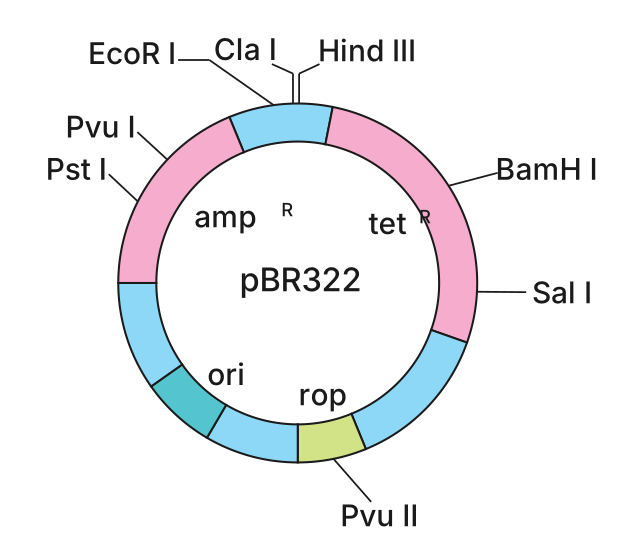
The ligation of foreign DNA is carried out at a restriction site located within one of the two antibiotic resistance genes. For example, in the well-known E. coli cloning vector pBR322, the BamH I restriction site lies inside the tetracycline resistance gene (tet<sup>R</sup>). When foreign DNA is inserted at this site, the tetracycline resistance gene is disrupted and becomes non-functional. The recombinant plasmid still retains its ampicillin resistance gene (amp<sup>R</sup>), so transformants can be selected by plating on ampicillin-containing medium. These transformants are then transferred to a medium containing tetracycline. Recombinants (with the insert) will grow on ampicillin but not on tetracycline, while non-recombinants (without the insert) will grow on both antibiotics.
This method of selection — using inactivation of one antibiotic resistance gene — is cumbersome because it requires plating on two different antibiotic media simultaneously.
To overcome this, alternative selectable markers have been developed that rely on a colour-based assay. In this system, the foreign DNA is inserted within the coding sequence of the enzyme β-galactosidase. This insertion inactivates the gene — a phenomenon called insertional inactivation. When a chromogenic substrate is present, bacterial colonies containing the intact β-galactosidase gene (no insert) turn blue. Colonies where the gene has been disrupted by an insert (recombinants) remain colourless. This allows direct visual identification of recombinants on a single plate.
(iv) Vectors for cloning genes in plants and animals
Nature has already perfected the art of delivering genes into eukaryotic cells. Bacteria and viruses have been doing this for ages to force host cells to produce what the pathogen needs. …
Drawn by us to help you understand the concept clearly, and verified to make sure it's accurate. For exams, practice from your textbook's own diagram.
The figure is a circular map of the plasmid pBR322, drawn as a closed loop. The circle represents the double-stranded DNA molecule. Around the circumference, specific restriction sites are marked as short tick marks labelled with the enzyme names: Hind III, EcoR I, BamH I, Sal I, Pvu II, Pst I, and Cla I. Each of these is a single recognition sequence — a key design feature because a vector should have only one cut site per restriction enzyme to avoid fragmenting the plasmid during cloning.
Inside the circle, three functional regions are labelled. The ori (origin of replication) is shown as a distinct segment; this is where DNA replication begins inside the host cell. The ampR and tetR genes are drawn as arcs along the plasmid, each encoding resistance to ampicillin and tetracycline respectively. These are the selectable markers. The rop gene is also indicated, positioned near the ori; it codes for proteins that regulate plasmid copy number.
The layout teaches a central principle of vector design. Notice that the restriction sites are deliberately placed within the antibiotic resistance genes. For example, BamH I lies inside the tetR gene, and Pst I lies inside the ampR gene. This allows insertional inactivation: when foreign DNA is ligated into a restriction site inside a resistance gene, that gene is disrupted and no longer functional. So a recombinant plasmid (one carrying an insert) will lose resistance to that particular antibiotic, while the other resistance gene remains intact. This makes selection possible — transformants are first selected on ampicillin medium (if the insert is in tetR), then replica-plated onto tetracycline medium. Recombinants grow on ampicillin but not on tetracycline; non-recombinants grow on both. …