Biology · Ch 5 — Molecular Basis of Inheritance
Structure of Polynucleotide Chain
Structure of Polynucleotide Chain
A polynucleotide chain is the fundamental chemical unit of both DNA and RNA. To understand how this chain stores genetic information, you must first know what a single building block — a nucleotide — looks like, and then how these blocks are linked together.
The Nucleotide: The Building Block
Every nucleotide has three components:
- A nitrogenous base — a nitrogen-containing ring compound.
- A pentose sugar — a five-carbon sugar. In RNA, this sugar is ribose; in DNA, it is deoxyribose (which lacks one oxygen atom on the 2' carbon).
- A phosphate group — derived from phosphoric acid.
Nitrogenous Bases: Purines and Pyrimidines
The nitrogenous bases are of two types:
- Purines — double-ringed structures: Adenine (A) and Guanine (G).
- Pyrimidines — single-ringed structures: Cytosine (C), Thymine (T), and Uracil (U).
Cytosine is common to both DNA and RNA. Thymine is found only in DNA. Uracil is found in RNA in place of thymine. (Thymine is chemically 5-methyl uracil.)
From Base to Nucleoside to Nucleotide
A nitrogenous base is attached to the 1' carbon of the pentose sugar through an N-glycosidic linkage. This base + sugar unit is called a nucleoside.
Examples of nucleosides:
- Adenosine (base A + ribose) or deoxyadenosine (base A + deoxyribose)
- Guanosine or deoxyguanosine
- Cytidine or deoxycytidine
- Uridine (RNA) or deoxythymidine (DNA)
When a phosphate group is attached to the 5' carbon of the nucleoside through a phosphoester linkage, the unit becomes a nucleotide (or deoxynucleotide, depending on the sugar).
Forming the Polynucleotide Chain
Two nucleotides are joined by a 3'-5' phosphodiester linkage — the phosphate group attached to the 5' carbon of one nucleotide bonds with the 3' carbon of the next nucleotide's sugar. This forms a dinucleotide. Repeating this process creates a long polynucleotide chain.
The chain has a clear direction:
- One end has a free phosphate group at the 5' carbon — this is the 5' end.
- The other end has a free –OH group at the 3' carbon — this is the 3' end.
The backbone of the chain is made of alternating sugar and phosphate groups. The nitrogenous bases project out from this backbone, like side branches.
Special Features of RNA
In RNA, every nucleotide residue has an additional –OH group at the 2' position of the ribose sugar. Also, RNA uses uracil instead of thymine.
Historical Context: The Discovery of DNA
DNA was first identified as an acidic substance in the nucleus by Friedrich Meischer in 1869. He named it 'nuclein'. However, isolating such a long polymer intact was technically difficult, so the structure of DNA remained unknown for decades.
The Watson-Crick Double Helix Model (1953)
James Watson and Francis Crick, using X-ray diffraction data from Maurice Wilkins and Rosalind Franklin, proposed the double helix model for DNA. A key feature was base pairing between the two strands. This was supported by Erwin Chargaff's observation: in any double-stranded DNA, the amount of adenine equals the amount of thymine, and the amount of guanine equals the amount of cytosine (A = T and G = C). The ratios A/T and G/C are constant and equal to one.
Salient Features of the Double Helix
- Two polynucleotide chains — the sugar-phosphate backbone is on the outside, and the bases project inside the helix.
- Anti-parallel polarity — one strand runs 5' → 3', and the other runs 3' → 5'.
- Base pairing via hydrogen bonds:
- Adenine (A) pairs with Thymine (T) using two hydrogen bonds.
- Guanine (G) pairs with Cytosine (C) using three hydrogen bonds.
- A purine always pairs with a pyrimidine, keeping the distance between the two strands uniform.
- Right-handed coiling — the helix twists to the right.
- Pitch (one complete turn) = 3.4 nm (nanometres).
- Each turn contains roughly 10 base pairs.
- Distance between adjacent base pairs = 0.34 nm.
- Base stacking — the flat planes of the base pairs stack on top of each other. This stacking, along with hydrogen bonds, gives the helix its stability.
The Key Consequence: Complementarity and Genetic Implication …
Drawn by us to help you understand the concept clearly, and verified to make sure it's accurate. For exams, practice from your textbook's own diagram.
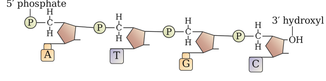
Figure 5.1 shows a short stretch of a single polynucleotide chain — the basic backbone of DNA or RNA. The chain is drawn as a vertical strand, with the sugar-phosphate backbone running along one side and the nitrogenous bases sticking out to the other side like side branches.
At the top of the chain, the 5' end is labelled: the sugar's 5' carbon carries a free phosphate group (shown as –PO₄). At the bottom, the 3' end is labelled: the sugar's 3' carbon has a free hydroxyl group (–OH). This polarity — 5' to 3' — is fundamental to how nucleotides are joined and how the chain grows.
Each repeating unit is a nucleotide, composed of three parts: a pentose sugar (a pentagon), a phosphate group (a circle labelled P), and a nitrogenous base (a flat rectangle or shape labelled A, G, C, T, or U). The sugar and phosphate alternate to form the backbone: the phosphate of one nucleotide is linked to the 3' carbon of the previous sugar via a 3'–5' phosphodiester bond (shown as a connecting line between the 3'–OH of one sugar and the phosphate of the next). The bases project outward from the 1' carbon of each sugar, attached by a N-glycosidic linkage.
The figure makes clear that the backbone is continuous and repeating (sugar–phosphate–sugar–phosphate), while the bases are not part of the backbone — they are side groups that will later face inward in a double helix. The free 5'–phosphate and free 3'–OH at the ends are the key landmarks that define the directionality of the chain. …
Drawn by us to help you understand the concept clearly, and verified to make sure it's accurate. For exams, practice from your textbook's own diagram.
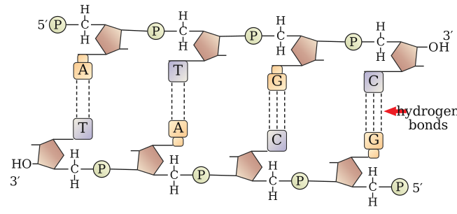
Figure 5.2 is a schematic of a short segment of double-stranded DNA, drawn to show the key structural features that Watson and Crick proposed. It is not a photograph or a detailed chemical diagram; it is a simplified, stylised representation meant to make the antiparallel nature and base-pairing rules visually clear.
The figure shows two polynucleotide chains running in opposite directions. Each chain is drawn as a ribbon or backbone, with the sugar-phosphate backbone on the outside and the nitrogenous bases projecting inward toward each other. The backbones are labelled with their directionality: one strand has its 5' end at the top and its 3' end at the bottom; the other strand runs 3' to 5' from top to bottom. This antiparallel arrangement is a central point of the figure.
Between the two backbones, the bases are paired. The figure uses a standard colour or shape code (often circles or rectangles) to distinguish the four bases: adenine (A), thymine (T), guanine (G), and cytosine (C). Each base pair is shown connected by dashed lines representing hydrogen bonds. A is always paired with T, and the figure indicates two hydrogen bonds between them. G is always paired with C, with three hydrogen bonds shown. This pairing is the basis of Chargaff's rule and the complementarity of the two strands.
The figure also makes clear that a purine (A or G, which are larger, two-ring structures) always pairs with a pyrimidine (T or C, which are smaller, single-ring structures). This keeps the distance between the two backbones nearly constant along the length of the helix — a point the textbook emphasises. The bases are stacked flat, one above the other, though the figure typically shows only a few base pairs in a row to illustrate the pattern. …
Drawn by us to help you understand the concept clearly, and verified to make sure it's accurate. For exams, practice from your textbook's own diagram.
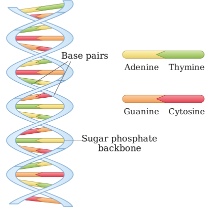
Figure 5.3 is a schematic representation of the DNA double helix as proposed by Watson and Crick. It shows two polynucleotide chains coiled around each other in a right-handed spiral. The backbone of each chain — made of alternating sugar and phosphate groups — is drawn as two smooth, ribbon-like strands running in opposite directions (antiparallel). The nitrogenous bases project inward from each backbone, like rungs of a twisted ladder, and pair specifically: adenine with thymine (two hydrogen bonds) and guanine with cytosine (three hydrogen bonds). The figure labels the pitch of the helix as 3.4 nm — the distance for one complete turn — and notes that each turn contains roughly 10 base pairs. The vertical spacing between adjacent base pairs is marked as 0.34 nm. The bases are shown stacked flat on top of one another, which contributes to the stability of the helix through base-stacking interactions. No arrows or additional labels appear beyond …