Biology · Ch 5 — Molecular Basis of Inheritance
The Genetic Material is DNA
The Genetic Material is DNA
The question of what actually carries hereditary information — the genetic material — was settled decisively by Alfred Hershey and Martha Chase in 1952. Before their experiment, both proteins and DNA were candidates, but proteins seemed more likely because they were more complex and diverse. Hershey and Chase provided the unequivocal proof that DNA, not protein, is the genetic material.
They worked with bacteriophages — viruses that infect bacteria. A bacteriophage works by attaching to a bacterial cell and injecting its genetic material inside. Once inside, the bacterium is tricked into treating the viral genetic material as its own and begins manufacturing more virus particles. The key question was: which component of the virus actually enters the bacterium — the protein coat or the DNA?
To find out, Hershey and Chase used radioactive isotopes as labels. They grew one batch of viruses in a medium containing radioactive phosphorus (phosphorus-32). DNA contains phosphorus, but protein does not, so only the viral DNA became radioactive. They grew another batch in a medium containing radioactive sulfur (sulfur-35). Proteins contain sulfur, but DNA does not, so only the viral protein coat became radioactive.
These radioactively labelled phages were then allowed to infect E. coli bacteria. After infection had begun, the researchers used a blender to agitate the mixture and strip the viral coats off the bacterial cells. They then separated the virus particles from the bacteria by spinning the mixture in a centrifuge.
The results were clear-cut:
- Bacteria infected with viruses that had radioactive DNA were themselves radioactive. This showed that DNA had entered the bacterial cells. …
Drawn by us to help you understand the concept clearly, and verified to make sure it's accurate. For exams, practice from your textbook's own diagram.
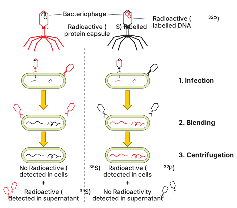
The figure is a multi-panel diagram that walks through the Hershey-Chase experiment step by step. It shows two parallel tracks, one for phages labelled with radioactive phosphorus ($^{32}\text{P}$) and one for phages labelled with radioactive sulphur ($^{35}\text{S}$). Each track begins with a bacteriophage particle — drawn as a small, insect-like structure with a head (containing the genetic material) and a tail — attached to the surface of an E. coli bacterium, shown as a larger oval.
In the first panel, the two types of labelled phages are shown separately: on the left, a phage whose DNA is marked with $^{32}\text{P}$ (often indicated by a star or dot inside the head); on the right, a phage whose protein coat is marked with $^{35}\text{S}$ (a star or dot on the head and tail surface). An arrow leads from each to the next panel, where the phage has injected its contents into the bacterium. The bacterial cell is now shown with the phage's head empty (or the radioactive label inside the bacterium for the $^{32}\text{P}$ track), while the phage's protein coat remains outside.
A blender symbol (often a small beaker or stirring icon) appears next, indicating that the mixture is agitated to shear off the empty phage coats from the bacterial surface. An arrow then leads to a centrifuge tube: the tube shows a pellet at the bottom (the heavier bacterial cells) and a supernatant above (the lighter phage coats). In the $^{32}\text{P}$ track, the pellet is labelled "radioactive" (or shown with a star), meaning the bacteria contain the radioactive DNA. In the $^{35}\text{S}$ track, the supernatant is labelled "radioactive" (or starred), meaning the radioactive protein stayed outside and did not enter the bacteria. …