Biology · Ch 1 — Sexual Reproduction in Flowering Plants
Embryo
Embryo
The embryo develops at the micropylar end of the embryo sac, precisely where the zygote is located. Most zygotes do not divide immediately after fertilisation — they wait until a certain amount of endosperm has formed. This is an adaptation: it ensures that the developing embryo has a guaranteed supply of nutrition from the very start.
Although mature seeds differ greatly in shape and size, the early stages of embryo development (called embryogeny) are remarkably similar in both dicotyledons and monocotyledons.
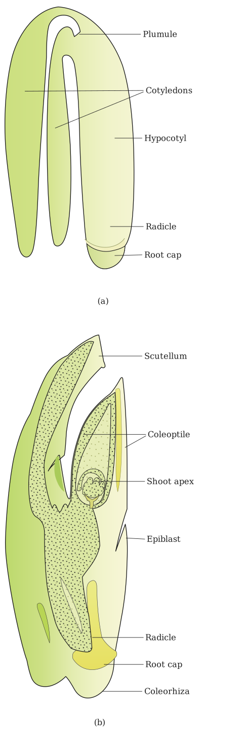
Embryogeny in a dicotyledonous embryo
The zygote first divides to form a proembryo. This proembryo then passes through a globular stage, followed by a heart-shaped stage, and finally develops into the mature embryo.
A typical dicotyledonous embryo consists of two main parts:
- Embryonal axis — the central stalk of the embryo.
- Two cotyledons — attached to the embryonal axis.
The embryonal axis is divided into two regions:
- Epicotyl — the portion above the level of the cotyledons. It terminates in the plumule (the stem tip).
- Hypocotyl — the cylindrical portion below the level of the cotyledons. It terminates at its lower end in the radicle (the root tip). The radicle is covered by a root cap.
Embryo of monocotyledons (especially grasses)
Monocot embryos possess only one cotyledon. In the grass family, this single cotyledon is called the scutellum. It is situated towards one side (lateral) of the embryonal axis.
The lower end of the embryonal axis has the radicle and root cap, both enclosed in an undifferentiated sheath called the coleorrhiza.
The portion of the embryonal axis above the level of attachment of the scutellum is the epicotyl. The epicotyl contains a shoot apex and a few leaf primordia, all enclosed in a hollow foliar structure called the coleoptile.
A quick way to observe these parts: soak seeds of wheat, maize, peas, chickpeas, or groundnut in water overnight. Split them open the next day and examine the embryo and seed structure.
Summary of key differences …
Drawn by us to help you understand the concept clearly, and verified to make sure it's accurate. For exams, practice from your textbook's own diagram.
Figure 1.14 places two mature embryos side by side so you can see the fundamental difference between dicots and monocots at a glance. Both panels are drawn as longitudinal sections, but the dicot is shown whole (a) while the monocot is explicitly labelled as an L.S. (b).
Panel (a) — a typical dicot embryo. The central structure is the embryonal axis, a vertical rod. Attached laterally at the same level are two fleshy cotyledons, one on each side. Above the point where the cotyledons join the axis is the epicotyl — the short stem portion that ends in the plumule (the shoot tip). Below the cotyledon attachment is the hypocotyl, the cylindrical lower part of the axis, which terminates in the radicle (the root tip). The radicle is capped by a root cap. So the layout is: plumule → epicotyl → (cotyledons) → hypocotyl → radicle + root cap.
Panel (b) — L.S. of a grass (monocot) embryo. Here there is only one cotyledon, called the scutellum, which sits to one side of the embryonal axis (lateral, not terminal). The axis itself is shorter and more compact. At the lower end, the radicle and its root cap are enclosed together inside a protective sheath called the coleorhiza — an undifferentiated covering that is unique to grasses. Above the level where the scutellum attaches, the epicotyl contains the shoot apex and a few tiny leaf primordia, all wrapped inside another protective sheath: the coleoptile. So the monocot embryo is built like a nested set: coleoptile encloses the shoot tip; coleorhiza encloses the root tip; and the scutellum sits alongside the axis, not around it. …