Chemistry · Ch 10 — Biomolecules
Structure of Nucleic Acids
Structure of Nucleic Acids
Building block 1: base + sugar → nucleoside
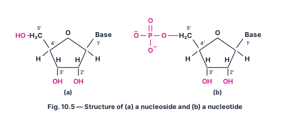
The first step in assembling a nucleic acid is attaching a base to the sugar. A base is linked to the position of the pentose sugar through a β-N-glycosidic linkage (the base's nitrogen bonds to the anomeric carbon of the sugar, with loss of water). The unit formed this way — base + sugar — is called a nucleoside.
(Fig. 10.5(a) shows this: the pentose ring with its – numbering, and the base attached at in place of the free that the isolated sugar carries there.)
Building block 2: nucleoside + phosphate → nucleotide
When a nucleoside is further linked to phosphoric acid through a phosphoester bond at the -position of the sugar, the resulting unit is called a nucleotide.
(Fig. 10.5(b) shows the completed nucleotide: a phosphate group, , esterified onto the - of the sugar, with the base still attached at .)
Drawn by us to help you understand the concept clearly, and verified to make sure it's accurate. For exams, practice from your textbook's own diagram.
Figure 10.5: Structure of (a) a nucleoside and (b) a nucleotide
The figure presents two side-by-side molecular structures that build the fundamental units of nucleic acids (DNA and RNA).
Panel (a) – A nucleoside
This is the simpler structure. It shows a pentose sugar drawn as a five-membered furanose ring. The ring has an oxygen atom at the top, and the carbon atoms are numbered through (the prime marks distinguish sugar carbons from base atoms). Attached to the carbon is a nitrogenous base (abbreviated as Base in the diagram). The sugar also carries:
- an group at and ,
- a group at .
Thus, a nucleoside is simply sugar + base.
Panel (b) – A nucleotide
This structure is identical to the nucleoside except that the group at is now esterified by a phosphate group (). The phosphate is drawn attached to the carbon. Hence, a nucleotide is sugar + base + phosphate.
Physical idea taught by the figure
The figure establishes the building-block relationship between nucleosides and nucleotides. A nucleotide is a nucleoside that has been phosphorylated at the carbon. This phosphate group is crucial because it allows nucleotides to link together via phosphodiester bonds (formed between the phosphate of one nucleotide and the of the next), creating the backbone of nucleic acid chains.
Key formula(s) developed with this figure
The textbook uses this figure to introduce the phosphodiester linkage that joins nucleotides. The general representation of a dinucleotide (two linked nucleotides) is:
where the bond forms between the carbon of one pentose sugar and the carbon of the next. In the chain, the repeating unit is:
…
A nucleotide is therefore the complete monomer unit of a nucleic acid — sugar in the middle, base hanging off , phosphate hanging off .
Linking nucleotides: the phosphodiester backbone
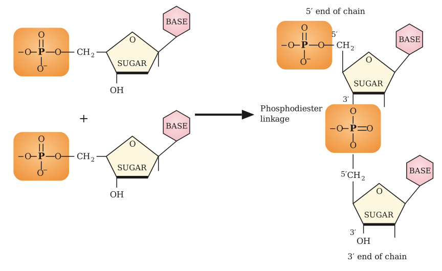
Nucleotides are joined to one another end-to-end by a phosphodiester linkage formed between the -carbon of one sugar and the -carbon of the next. Concretely: the free -phosphate of one nucleotide condenses with the free -OH of the sugar of the neighbouring nucleotide, releasing a molecule of water and leaving a phosphate group bridged (diester-linked) between the two sugars.
(Fig. 10.6 walks through exactly this: two separate nucleotides, each still carrying a free -phosphate and a free -OH, combine through a phosphodiester linkage to give a dinucleotide — a phosphate now bridges the -oxygen of the first sugar to the -oxygen of the second.)
Drawn by us to help you understand the concept clearly, and verified to make sure it's accurate. For exams, practice from your textbook's own diagram.
What the Figure Shows
The figure illustrates the formation of a dinucleotide — the simplest unit of a nucleic acid chain. It depicts two nucleotides linked together by a phosphodiester bond. Each nucleotide consists of three components: a pentose sugar (ribose in RNA, deoxyribose in DNA), a nitrogenous base (attached to the 1′ carbon of the sugar), and a phosphate group (attached to the 5′ carbon of the sugar).
In the diagram, the first nucleotide has its phosphate group at the 5′ carbon of its sugar. This phosphate forms a bridge to the 3′ carbon of the second nucleotide’s sugar. The linkage is specifically between the 5′-phosphate of one nucleotide and the 3′-hydroxyl (–OH) group of the next. This creates a 5′→3′ phosphodiester linkage, which is the backbone of all nucleic acids.
The bases (adenine, guanine, cytosine, thymine, or uracil) are shown projecting outward from each sugar, not participating in the backbone. The figure also labels the sugar–phosphate backbone and the bases as separate components, emphasizing that the repeating sugar–phosphate units form the structural framework, while the bases carry genetic information.
Physical Idea Taught
The figure teaches the directionality and connectivity of nucleic acid strands. The 5′→3′ linkage is fundamental because it determines how nucleotides polymerize. Each new nucleotide is added to the 3′ end of the growing chain, with its own 5′-phosphate forming a bond to the 3′-OH of the previous sugar. This creates a polar strand — one end has a free 5′-phosphate (the 5′ end), and the other has a free 3′-OH (the 3′ end).
The figure also introduces the concept of primary structure: the sequence of nucleotides along the chain. The repeating sugar–phosphate backbone is invariant, but the order of bases (A, T, G, C in DNA; A, U, G, C in RNA) encodes genetic information. This is the foundation for understanding DNA replication, transcription, and the double helix.
Key Formula and Symbol Explanation
The textbook develops the phosphodiester bond as a chemical linkage. The general reaction for joining two nucleotides is:
In terms of the backbone structure, the repeating unit is:
Where:
- 5′ and 3′ refer to the carbon atoms of the pentose sugar (ribose or deoxyribose). The prime (′) distinguishes these from atoms in the base.
- Phosphate is the group that bridges two sugars.
- Sugar is the pentose (CHO for deoxyribose, CHO for ribose). …
Repeating this linkage over and over builds a long chain — a polynucleotide — with the general repeating pattern:
with a base projecting from every sugar. Because the phosphodiester bridge always runs from a -carbon on one sugar to a -carbon on the next, the chain has a built-in sense of direction:
- one end of the chain retains a free -phosphate group — the end
- the other end retains a free -OH group — the end
This directionality is a defining feature of every nucleic acid strand, and it is what makes it meaningful to talk about the two strands of DNA running in opposite directions (below).
The exact sequence in which nucleotides (and hence bases) occur along this chain is called the primary structure of the nucleic acid — it is this sequence that ultimately encodes genetic information.
Secondary structure: the DNA double helix
Nucleic acids also have a well-defined secondary structure — the three-dimensional shape the polynucleotide chain(s) fold or coil into.
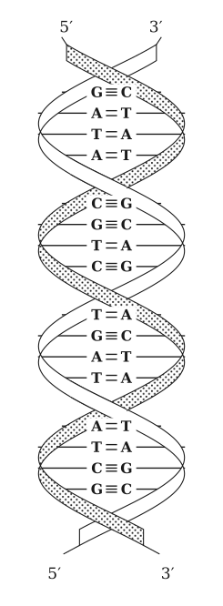
For DNA, James Watson and Francis Crick proposed the now-famous double helix model: two separate polynucleotide chains are wound around a common axis, coiling around each other, and are held together by hydrogen bonds formed between pairs of bases, one base projecting inward from each strand. Because these hydrogen bonds only form between specific, matching pairs of bases, the two strands of the helix are complementary to each other — the base sequence of one strand completely determines the base sequence of the other.
Base-pairing rule and why the strands are complementary
- Adenine (A) hydrogen-bonds specifically with Thymine (T) — a purine paired with a pyrimidine, held by two hydrogen bonds.
- Guanine (G) hydrogen-bonds specifically with Cytosine (C) — a purine paired with a pyrimidine, held by three hydrogen bonds.
- No other pairing (A–C, A–G, T–G, etc.) fits geometrically to form stable hydrogen bonds across the helix.
- Because only A↔T and G↔C pairs are allowed, the base at any position on one strand fixes the base that must sit opposite it on the other strand — the two strands carry the same information twice, written in complementary form. Knowing one strand's sequence lets you read off the other strand's sequence exactly. This complementarity is also the chemical basis for how DNA can be faithfully copied (see §10.5.3). …
Drawn by us to help you understand the concept clearly, and verified to make sure it's accurate. For exams, practice from your textbook's own diagram.
The figure depicts the double helix model of DNA, as proposed by Watson and Crick. Two sugar–phosphate backbones (shown as dotted ribbons) coil around a common axis in a right-handed spiral. The strands run in opposite directions — one is oriented downward, the other upward — making them antiparallel.
Between the backbones, horizontal rungs represent the complementary base pairs. Each rung consists of one purine (adenine or guanine) from one strand paired with one pyrimidine (thymine or cytosine) from the other. The pairing is specific:
- Adenine (A) pairs with thymine (T) via two hydrogen bonds ().
- Guanine (G) pairs with cytosine (C) via three hydrogen bonds ().
This complementary base pairing holds the two strands together and is the key to the molecule’s stability and its role in heredity.
Physical idea taught: The double helix is the secondary structure of DNA. The sequence of nucleotides along one strand (the primary structure) determines the complementary sequence on the other strand. The hydrogen bonds between specific base pairs allow the two strands to separate and serve as templates during replication and transcription.
Key formula(s) developed with this figure:
The number of hydrogen bonds in a DNA segment depends on the base composition. For a segment with adenine–thymine pairs and guanine–cytosine pairs, the total number of hydrogen bonds is:
where: …