Botany · Ch 5 — Cell: The Unit of Life
An Overview of Cell
An Overview of Cell
A quick tour of cell structure shows both how much cells have in common and how greatly they differ. The familiar examples from earlier observation — an onion peel and human cheek cells — make a good starting point.
The outer boundary and the nucleus
- The onion cell is a typical plant cell. Its outermost boundary is a distinct cell wall, and lying just inside the wall is the cell membrane.
- The human cheek cell has an outer membrane as its delimiting structure, but no cell wall.
- Inside each cell sits a dense, membrane-bound structure, the nucleus. The nucleus holds the chromosomes, which in turn carry the genetic material, DNA.
This single feature — whether or not the nucleus is enclosed in a membrane — divides all cells into two great groups:
- Eukaryotic cells have a membrane-bound nucleus.
- Prokaryotic cells lack a membrane-bound nucleus.
The cytoplasm
In both prokaryotic and eukaryotic cells, a semi-fluid matrix called the cytoplasm fills the volume of the cell. The cytoplasm is the main arena of cellular activity in plant and animal cells alike — it is where the many chemical reactions that keep the cell in the 'living state' take place.
Organelles
Besides the nucleus, eukaryotic cells contain other membrane-bound structures called organelles, including:
- the endoplasmic reticulum (ER)
- the golgi complex
- lysosomes
- mitochondria
- microbodies
- vacuoles
Prokaryotic cells lack these membrane-bound organelles.
Ribosomes are a special case: they are non-membrane-bound organelles present in all cells, both eukaryotic and prokaryotic. Within a cell, ribosomes occur not only in the cytoplasm but also inside two organelles — chloroplasts (in plants) and mitochondria — and on the rough ER. Animal cells also contain another non-membrane-bound organelle, the centrosome, which assists in cell division.
Variation in size, shape and number
Cells differ enormously in size, shape and activity. …
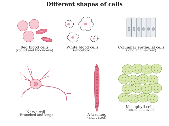
Drawn by us to help you understand the concept clearly, and verified to make sure it's accurate. For exams, practice from your textbook's own diagram.
This figure shows how differently shaped cells can be, even though every one of them is built from the same basic plan. Red blood cells are round and biconcave, shaped to carry oxygen efficiently. White blood cells are amoeboid, with no single fixed shape, letting them squeeze between other cells. Columnar epithelial cells are long and narrow, packed side by side to line surfaces such as the gut. A nerve cell is branched and unusually long, its shape suited to carrying electrical impulses over distance. A tracheid is an elongated, tapering plant cell that forms part of the water-conducting xylem tissue. Mesophyll cells are round to oval, packed tightly together inside a leaf, where their shape helps them car …