Zoology · Ch 4 — Excretory Products and their Elimination
Human Excretory System
Human Excretory System
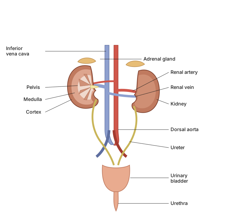
In humans, the excretory system is built around a small set of organs that together produce, carry, store and expel urine: a pair of kidneys, a pair of ureters, a single urinary bladder and a urethra.
The kidneys
The kidneys are the central organs of this system. They are reddish-brown, bean-shaped structures lying against the back inner wall of the abdominal cavity, positioned roughly between the last thoracic vertebra and the third lumbar vertebra. In an adult, each kidney is about 10 to 12 cm long, 5 to 7 cm wide and 2 to 3 cm thick, and weighs on average between 120 and 170 grams.
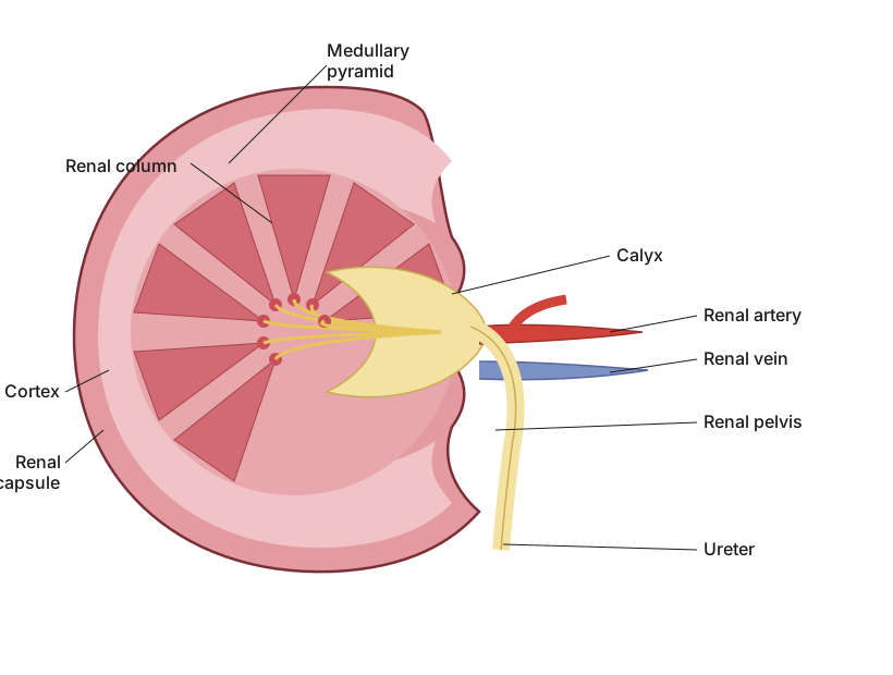
The inner, concave surface of each kidney carries a notch called the hilum. This is the gateway of the organ: the ureter leaves through it, and blood vessels and nerves enter through it. Just inside the hilum lies a broad, funnel-shaped chamber, the renal pelvis, whose finger-like extensions are called calyces.
Internal zones
- The whole kidney is wrapped in a tough outer capsule.
- Beneath the capsule the tissue is organised into two zones: an outer cortex and an inner medulla.
- The medulla is not uniform; it is broken into a few cone-shaped masses called medullary pyramids, whose tips project into the calyces.
- The cortical tissue does not stop at the boundary of the medulla -- it dips down between the pyramids as extensions known as renal columns or Columns of Bertini.
The nephron -- the functional unit
Each kidney contains close to one million tiny, complex tubular structures called nephrons. These are the true working units where urine is made. A nephron has two main parts: the glomerulus and the renal tubule.
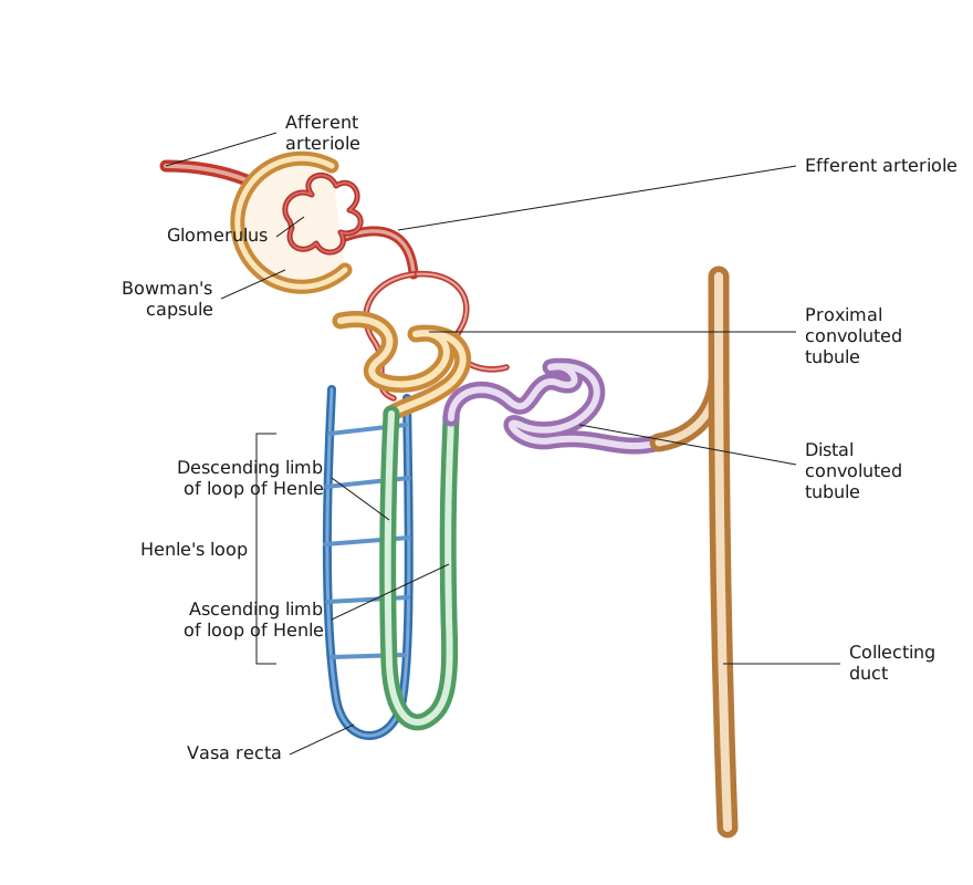
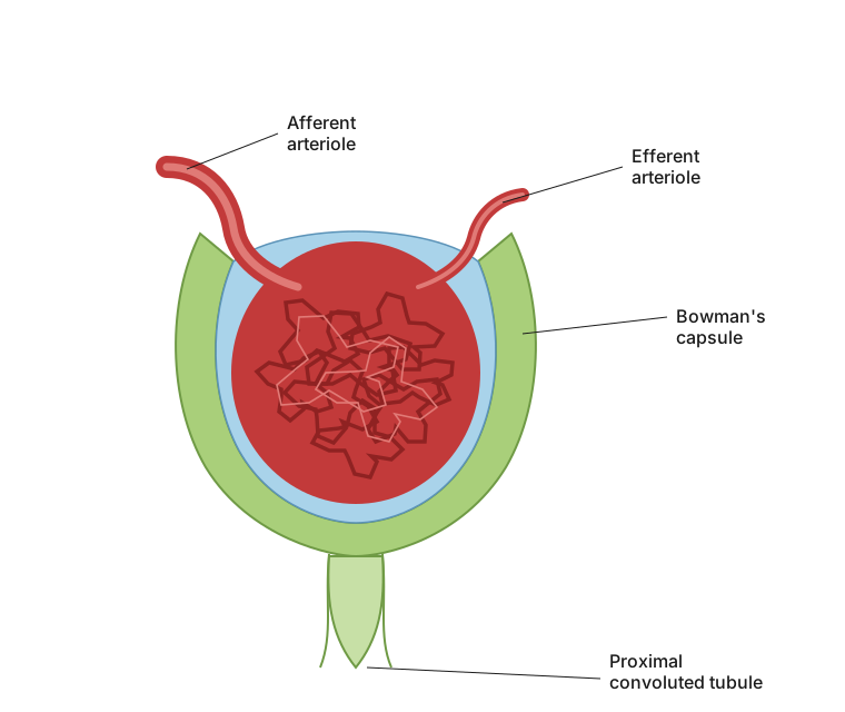
The glomerulus is a compact tuft of capillaries. It is supplied by the afferent arteriole, a fine branch of the renal artery, and the blood leaving it is drained away by the efferent arteriole.
The renal tubule begins as a double-walled, cup-shaped structure called Bowman's capsule, which wraps around the glomerulus. The glomerulus together with Bowman's capsule is known as the Malpighian body or renal corpuscle. From here the tubule runs on through several distinct stretches:
- Proximal convoluted tubule (PCT): a highly coiled network continuing from Bowman's capsule.
- Henle's loop: a hairpin-shaped segment with a descending limb going down and an ascending limb coming back up.
- Distal convoluted tubule (DCT): another highly coiled region continuing from the ascending limb.
- Collecting duct: the DCTs of many nephrons empty into these straight tubes. Many collecting ducts then converge and open into the renal pelvis, passing through the medullary pyramids at the calyces.
Where the nephron sits, and two types of nephrons
The Malpighian corpuscle, the PCT and the DCT all lie in the cortex, while the loop of Henle dips down into the medulla. How far the loop reaches divides nephrons into two kinds:
- Cortical nephrons: the majority. Their loop of Henle is short and barely enters the medulla. …
Drawn by us to help you understand the concept clearly, and verified to make sure it's accurate. For exams, practice from your textbook's own diagram.
This figure lays out the complete human excretory system so you can trace the path that urine takes from where it is made to where it leaves the body. Look first for the pair of bean-shaped kidneys set against the back inner wall of the abdomen, and from each kidney follow the tube running downward that carries urine away, the ureter. Both ureters lead into a single muscular sac, the urinary bladder, which stores urine until it is passed out through the urethra. Reading the figure in this order shows that the system works like a simple assembly line: two organs tha …
Drawn by us to help you understand the concept clearly, and verified to make sure it's accurate. For exams, practice from your textbook's own diagram.
This figure opens up a single kidney so you can study its inside rather than its outer bean shape. Notice the notch on the inner concave edge, the hilum, which acts as the organ's gateway where the ureter leaves and where blood vessels and nerves enter. Just within it, spot the broad funnel-shaped renal pelvis together with its finger-like branches, the calyces. The cut surface also reveals two clear tissue zones: an outer cortex and an inner medulla. The medulla is not smooth but is broken into cone-shaped medullary pyramids whose tips point into the calyces, while strips of cortical tis …
Drawn by us to help you understand the concept clearly, and verified to make sure it's accurate. For exams, practice from your textbook's own diagram.
This figure follows a single nephron from where blood enters it to where the filtrate leaves it, showing both the blood vessels and the tubule side by side. Start at the afferent arteriole feeding into the glomerulus, the tuft of capillaries cupped inside Bowman's capsule; blood leaves again through the efferent arteriole. From Bowman's capsule, follow the renal tubule as it winds into the proximal convoluted tubule, then straightens into the hairpin-shaped loop of Henle with its descending and ascending limbs, then coils again as the distal convoluted tubule before draining into the collecting duct. Running alongside the loop of Henle is the vasa recta, the U-shaped capillary network that stays closely paired with the tubule. Reading the figure this way shows the complete path blood and filtrate tak …
Drawn by us to help you understand the concept clearly, and verified to make sure it's accurate. For exams, practice from your textbook's own diagram.
This figure zooms in on the tiny filtering unit where blood is first sieved, letting you see how ultrafiltration actually happens. Identify the compact ball of capillaries, the glomerulus, cupped inside the surrounding Bowman's capsule. The figure highlights the three layers that plasma must be pushed across to become filtrate: the inner lining of the glomerular capillaries, a basement membrane in the middle, and the epithelium of Bowman's capsule. Look closely at these epithelial cells, the podocytes, arranged so that narrow filtration slits are left between them. Because these membranes are so fine, almost every part of t …