Zoology · Ch 7 — Human Reproduction
The Male Reproductive System
The Male Reproductive System
The male reproductive system is located in the pelvis region. It is a system built for the production, storage, and transport of sperm, as well as the delivery of these gametes into the female reproductive tract.
The system consists of four main components: a pair of testes, a set of accessory ducts, several accessory glands, and the external genitalia.
The Testes (Primary Sex Organs)
The testes are the primary male reproductive organs. They are a pair, each oval-shaped, and are located outside the abdominal cavity within a pouch of skin called the scrotum. This placement is critical — the scrotum maintains the testes at a temperature 2–3°C lower than the core body temperature, which is essential for proper sperm formation (spermatogenesis).
Each testis is about 4–5 cm in length and 2–3 cm in width. Internally, it is divided into about 250 compartments called testicular lobules. Each lobule contains one to three highly coiled seminiferous tubules. These tubules are the actual sites of sperm production.
The seminiferous tubules are lined by two types of cells:
- Male germ cells (spermatogonia): These are the cells that undergo meiotic divisions to eventually form spermatozoa.
- Sertoli cells (nurse cells): These provide nourishment to the developing germ cells.
In the spaces between the seminiferous tubules (the interstitial spaces), you find interstitial cells or Leydig cells. These cells synthesise and secrete the male sex hormone, testosterone.
Accessory Ducts
The ducts form a passageway for the transport of sperm from the testes to the outside. The sequence is as follows:
- Rete testis: Sperm from the seminiferous tubules are first moved into this network of channels within the testis.
- Vasa efferentia: From the rete testis, the sperm pass through these small ducts.
- Epididymis: This is a long, coiled tube that lies along the posterior surface of each testis. It stores the sperm and allows them to mature and become motile.
- Vas deferens: This is a thick-walled, muscular tube that ascends from the scrotum into the abdominal cavity. It loops over the urinary bladder and joins the duct of the seminal vesicle to form the ejaculatory duct.
- Ejaculatory duct: This short duct passes through the prostate gland and opens into the urethra.
- Urethra: This is the common passage for both urine and semen. It runs through the penis and opens to the outside at the external urethral orifice.
Accessory Glands
These glands secrete fluids that nourish, protect, and activate the sperm. The secretions of all three glands together form the bulk of the semen.
- Seminal vesicles: A pair of glands located behind the urinary bladder. They secrete a viscous, alkaline fluid rich in fructose (an energy source for sperm), calcium, and certain clotting proteins.
- Prostate gland: A single, large gland that surrounds the urethra just below the urinary bladder. It secretes a thin, milky, alkaline fluid that helps neutralise the acidity of the female vagina.
- Bulbourethral glands (Cowper's glands): A pair of small glands located on either side of the urethra below the prostate. They secrete a mucus-like fluid that lubricates the urethra and the tip of the penis before ejaculation.
External Genitalia …
Drawn by us to help you understand the concept clearly, and verified to make sure it's accurate. For exams, practice from your textbook's own diagram.
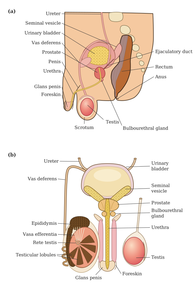
This figure is a two-panel diagram that serves as the primary visual guide to the male reproductive system. Panel (a) gives you a mid-sagittal (side) cutaway of the male pelvis. This view shows how the reproductive organs sit relative to the urinary bladder and the urethra. You see the testis hanging outside the body cavity inside the scrotum, which keeps it cooler than core body temperature. Behind the testis lies the coiled epididymis, the site where sperm mature and are stored. From the epididymis, the vas deferens runs upward, loops over the ureter, and passes behind the urinary bladder. Along its path, it is joined by ducts from the seminal vesicle (a gland that secretes a fructose-rich fluid) and then passes through the prostate gland, which surrounds the urethra just below the bladder. Further down, near the base of the penis, you see the small bulbourethral gland (also called Cowper's gland). The urethra is the common passage for both urine and semen, running through the length of the penis, which is the external genital organ.
Panel (b) is a frontal view with one testis cut open to reveal its internal architecture. Inside the testis, you see it is divided into about 250 testicular lobules by connective tissue septa. Each lobule contains one to three highly coiled seminiferous tubules — these are the actual sites of sperm production (spermatogenesis). The seminiferous tubules converge toward the back of the testis to form a network of channels called the rete testis. From the rete testis, 15–20 small ducts called vasa efferentia pierce the testis wall and enter the head of the epididymis. The epididymis is shown as a long, coiled tube that continues into the vas deferens, which then ascends into the abdomen.
The key relationship to grasp: sperm are made in the seminiferous tubules → move into the rete testis → exit via vasa efferentia → enter the epididymis for storage and maturation → travel through the vas deferens during ejaculation. …
Drawn by us to help you understand the concept clearly, and verified to make sure it's accurate. For exams, practice from your textbook's own diagram.
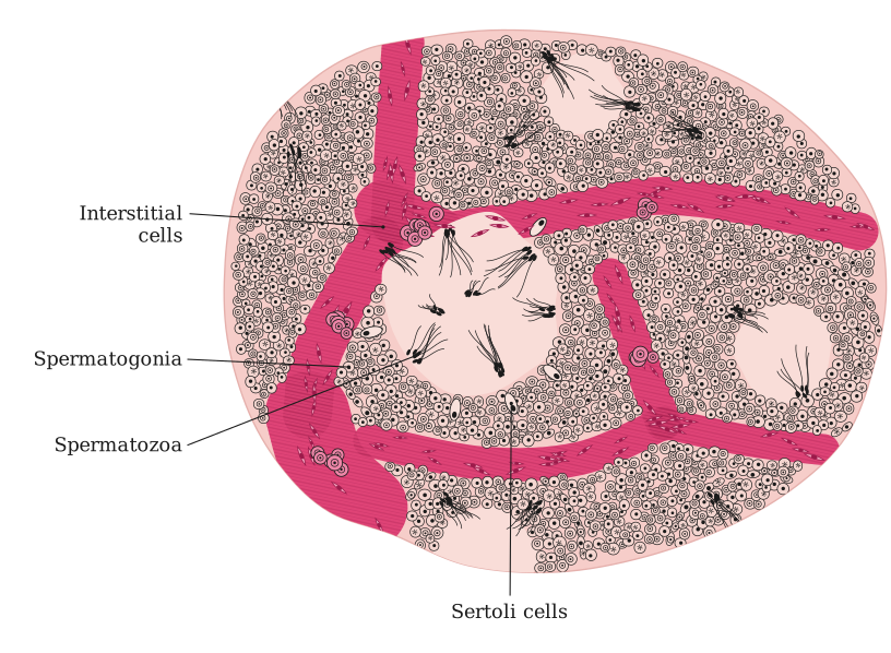
The figure is a transverse (cross) section of a single seminiferous tubule, cut perpendicular to its length so you see it as a circle or oval. The tubule is the functional unit of the testis where sperm are produced.
What the wall shows: The inner lining of the tubule is not a simple epithelium — it is a stratified (layered) arrangement of cells. The outermost layer, closest to the basement membrane, consists of spermatogonia (the male germ cells). These are the stem cells that divide by mitosis to maintain their own population and to produce primary spermatocytes. Scattered among the spermatogonia are Sertoli cells (also called sustentacular cells). These are tall, columnar cells that extend from the basement membrane all the way to the lumen. They provide nourishment, support, and protection to the developing sperm cells, and they also secrete fluid and hormones (like inhibin).
Moving inward toward the lumen: As spermatogonia undergo meiosis and differentiation, the developing cells — primary spermatocytes, secondary spermatocytes, spermatids, and finally spermatozoa — are seen in successive layers closer to the central cavity. The most mature sperm cells are found right at the lumen (the hollow centre of the tubule), with their heads embedded in the Sertoli cells and their tails projecting into the lumen. This arrangement shows the progressive maturation of sperm from the periphery to the centre.
Outside the tubule: Between adjacent seminiferous tubules lies the interstitial space. This space contains Leydig cells (interstitial cells) and blood vessels. Leydig cells are endocrine cells that produce the male sex hormone testosterone under the influence of luteinising hormone (LH) from the pituitary. The blood vessels supply oxygen and nutrients and carry away hormones and waste. …