Biology · Ch 17 — Locomotion and Movement
Skeletal System
Skeletal System
Working alongside the muscles is the skeletal system -- the body's supporting scaffold, built chiefly of bones with a handful of cartilages filling in at the flexible points. Its contribution to movement is easy to underestimate until you imagine living without it: no jaw bones would mean no chewing, and no limb bones would mean no walking at all.
Bone and cartilage as connective tissues
Both bone and cartilage are specialised connective tissues, but they differ in the nature of their matrix:
- Bone's matrix is rock-solid, a hardness that comes directly from the calcium salts deposited within it.
- Cartilage, by contrast, has some give to it -- a flexibility owed to the chondroitin salts that make up its matrix instead.
In a human being the skeletal system is made up of 206 bones together with a few cartilages. These are organised into two main divisions: the axial skeleton and the appendicular skeleton.
Axial skeleton
The axial skeleton consists of 80 bones arranged along the central axis of the body. Its parts are the skull, vertebral column, sternum and ribs.
The skull
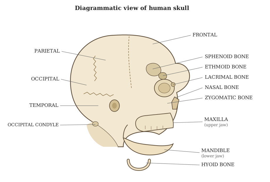
The skull is built from two sets of bones — cranial and facial — which together total 22 bones.
- Cranial bones number 8. They form the hard, protective outer case, the cranium, that encloses the brain.
- Facial bones number 14 and make up the front part of the skull.
Related to the skull region:
- A single U-shaped bone, the hyoid, lies at the base of the buccal cavity (mouth).
- Each middle ear contains three tiny bones — the malleus, incus and stapes — together called the ear ossicles (six in the two ears).
The skull rests on the vertebral column: it joins the top of the column through two occipital condyles, which is why the human skull is described as dicondylic.
Vertebral column
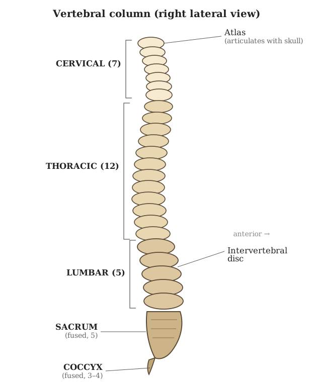
The vertebral column is formed by 26 serially arranged units called vertebrae and lies along the dorsal (back) side. It runs from the base of the skull and forms the main framework of the trunk.
- Each vertebra has a central hollow, the neural canal, through which the spinal cord passes.
- The first vertebra is the atlas, and it articulates with the occipital condyles of the skull.
Starting from the skull, the column is divided into regions:
- Cervical — 7 vertebrae
- Thoracic — 12 vertebrae
- Lumbar — 5 vertebrae
- Sacral — 1 (fused)
- Coccygeal — 1 (fused)
Interestingly, the number of cervical vertebrae is seven in almost all mammals, humans included. The vertebral column protects the spinal cord, supports the head, and provides attachment for the ribs and the muscles of the back.
Sternum and ribs
The sternum is a flat bone lying on the ventral midline of the thorax.
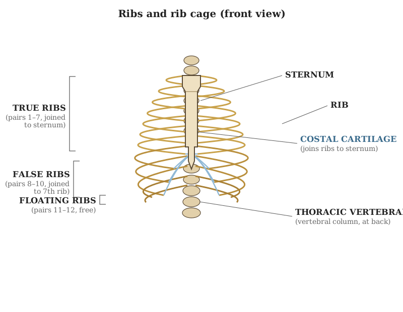
There are 12 pairs of ribs. Each rib is a thin, flat bone attached dorsally to the vertebral column and ventrally to the sternum. Because a rib has two articulation surfaces on its dorsal end, it is described as bicephalic. The ribs fall into three groups:
- True ribs — the first 7 pairs. Dorsally they attach to the thoracic vertebrae; ventrally they connect to the sternum through hyaline cartilage.
- False (vertebrochondral) ribs — the 8th, 9th and 10th pairs. These do not join the sternum directly; instead each joins the seventh rib through hyaline cartilage.
- Floating ribs — the last 2 pairs (11th and 12th). These are not connected ventrally at all.
Together, the thoracic vertebrae, the ribs and the sternum form the rib cage.
Appendicular skeleton
The appendicular skeleton is made of the limb bones together with their girdles. Each limb contains 30 bones.
Bones of the forelimb (hand/arm)
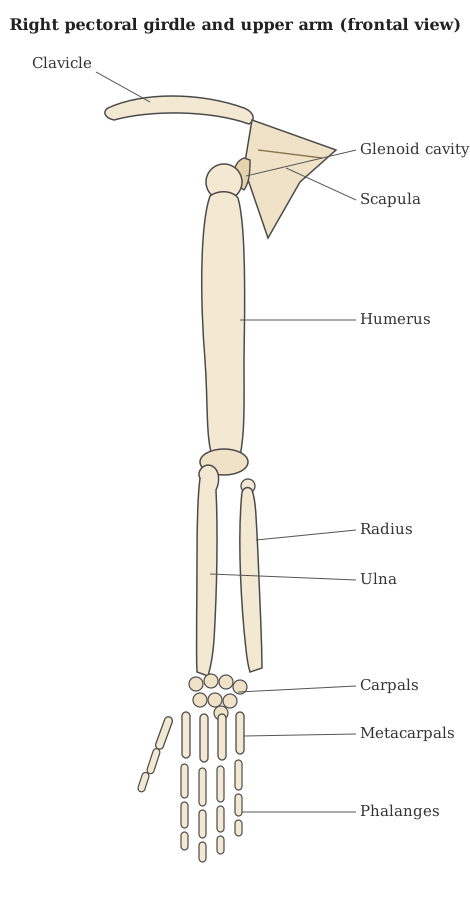
- Humerus — the upper arm bone
- Radius and ulna — the two bones of the forearm
- Carpals — the wrist bones, 8 in number
- Metacarpals — the palm bones, 5 in number
- Phalanges — the finger bones (digits), 14 in number
Bones of the hind limb (leg)
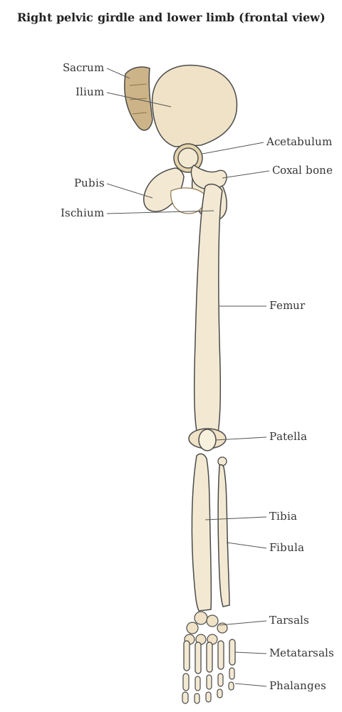
- Femur — the thigh bone, which is the longest bone of the body
- Tibia and fibula — the two bones of the lower leg
- Tarsals — the ankle bones, 7 in number
- Metatarsals — 5 in number
- Phalanges — the toe bones (digits), 14 in number
- Patella — a cup-shaped bone covering the knee on its ventral side, commonly called the knee cap
Girdles
Two girdles anchor the limbs to the main skeleton -- the pectoral girdle for the arms and the pelvic girdle for the legs -- and each is built from a matching pair of halves, one on the left and one on the right.
Pectoral girdle. Each side pairs a scapula with a clavicle. …
Drawn by us to help you understand the concept clearly, and verified to make sure it's accurate. For exams, practice from your NCERT textbook's own diagram.
This diagram shows the bones that make up the human skull, which has two main parts. The first is the cranium, a bony case of eight bones that surrounds and protects the brain: the frontal bone at the forehead, two parietal bones forming the top and sides, two temporal bones near the ears, the occipital bone at the back, and the sphenoid and ethmoid bones deeper inside. At the base of the occipital bone are two small projections, the occipital condyles, which are where the skull rests on the topmost vertebra of the neck. The second part is the group of fourteen facial bones that shape the face, including the maxillae of the upper jaw, the small zygomatic (cheek) bones, the paired nasal bones forming the bridge of the nose, and the tiny lacrimal bones at the inner corner of each eye socket. Note that the lower jaw, the mandible, is the only freely movable bone of the skull and lets us chew and speak. A separate U-shaped bone, the hyoid, sits below the mandible at the base of the mouth -- not par …
Drawn by us to help you understand the concept clearly, and verified to make sure it's accurate. For exams, practice from your NCERT textbook's own diagram.
Seen from the side, this figure shows the vertebral column, or backbone, which in an adult is made of 26 bones stacked one above another. From top to bottom there are 7 cervical vertebrae in the neck, 12 thoracic vertebrae in the chest region, and 5 lumbar vertebrae in the lower back. Below these lie the sacrum -- itself a single fused bone, commonly described as formed from five fused vertebrae -- and finally the coccyx or tail bone, another single fused bone usually cited as three to four fused vertebrae (sources vary slightly on the exact count). The topmost cervical vertebra, the atlas, articulates with the occipital condyles at the base of the skull -- the joint that, among its other roles, is commonly credited with letting us nod the head. Between one vertebra and the next sits a cushioning pad of cartilage, the intervertebral disc, which lets the column flex slightly at every joint while absorbing shock. Running through the column …
Drawn by us to help you understand the concept clearly, and verified to make sure it's accurate. For exams, practice from your NCERT textbook's own diagram.
This figure shows the rib cage, the bony basket that guards the heart and lungs. There are 12 pairs of ribs, and each rib is joined at the back to a thoracic vertebra of the backbone. In front, the ribs differ in how they connect. The first seven pairs are the true ribs; each joins directly to the breastbone, or sternum, through a strip of cartilage. Pairs eight, nine and ten are the false ribs; instead of reaching the sternum on their own, they attach to the cartilage of the seventh rib above them. The last two pairs, eleven and twelve, are the floating ribs, because they are not connected to …
Drawn by us to help you understand the concept clearly, and verified to make sure it's accurate. For exams, practice from your NCERT textbook's own diagram.
This front view shows the right shoulder region and the bones of the arm. The pectoral girdle, which anchors the arm to the trunk, is made of two bones: the clavicle, or collar bone, in front, and the scapula, or shoulder blade, behind. The scapula carries a shallow socket called the glenoid cavity, into which the arm bone fits to form the shoulder joint. Moving down the limb, the upper arm contains a single long bone, the humerus. Below the elbow, the forearm has two bones lying side by side, the radius and the ulna. These lead to the wrist, made of small carpal bones, …
Drawn by us to help you understand the concept clearly, and verified to make sure it's accurate. For exams, practice from your NCERT textbook's own diagram.
This front view shows the right hip region and the bones of the leg. The pelvic girdle attaches the lower limb to the trunk and, together with the sacrum at the base of the spine, forms the bowl-shaped pelvis. Each hip bone -- properly called the coxal bone -- is actually three bones fused together, the ilium, the ischium and the pubis, and they meet at a deep cup called the acetabulum, which forms the socket of the hip joint. Fitting into this socket is the femur, the long, strong thigh bone. At the knee sits a small bone, the patella or knee cap. Below the knee the lower leg has two bones, the larger tibia and the slender fibula. These lead down to the ankle, fo …