Biology · Ch 10 — Biotechnology and Its Applications
Genetically Engineered Insulin
Genetically Engineered Insulin
Insulin is a protein hormone essential for managing blood sugar levels. Patients with adult-onset diabetes must take insulin at regular intervals. Before genetic engineering, the only source of insulin was the pancreas of slaughtered cattle and pigs. This animal-derived insulin was not identical to human insulin, and in some patients it triggered allergic reactions or other immune responses because the body recognised it as a foreign protein.
The ideal solution would be to have a bacterium that could be engineered to produce human insulin. Bacteria can be grown in large quantities, making the process simple and scalable. However, insulin cannot be taken orally because it is a protein — digestive enzymes in the stomach and intestine would break it down before it could enter the bloodstream. It must therefore be injected.
Structure of insulin
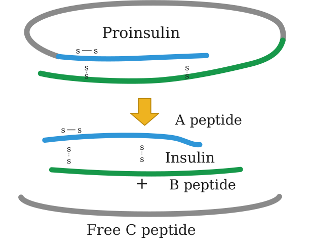
Insulin consists of two short polypeptide chains: chain A and chain B. These two chains are linked together by disulphide bridges (as shown in Figure 10.3 of the NCERT textbook).
The pro-hormone problem
In mammals, including humans, insulin is not synthesised directly in its active form. It is first made as a pro-hormone — a longer, inactive precursor that contains an extra stretch of amino acids called the C peptide. This C peptide is not present in the mature insulin molecule; it is removed during processing to yield the functional hormone. This is similar to how a pro-enzyme must be cleaved to become active.
This posed the main challenge for producing insulin using recombinant DNA technology: how to get the insulin polypeptide chains to assemble correctly into the mature, active form.
The breakthrough (1983, Eli Lilly) …
Drawn by us to help you understand the concept clearly, and verified to make sure it's accurate. For exams, practice from your NCERT textbook's own diagram.
Figure 10.3 is a simplified schematic, laid out top-to-bottom, that shows the two-stage life of insulin inside a human cell.
At the top, an oval outline labelled Proinsulin encloses a single, long, folded polypeptide chain. Even at this stage, disulphide bridges (S–S bonds) are already shown holding the chain's two future halves together.
A single downward arrow leads from the proinsulin oval to the structures below it. (The arrow itself carries no printed word — "maturation" is the term used in the figure's own caption below the image, not a label drawn on the arrow.)
Below the arrow, two separate structures are shown:
- Insulin — the two short chains that remain linked by disulphide bridges, labelled A peptide and, joined with a + sign, B peptide.
- Free C peptide — drawn as its own separate oval outline, positioned at the very bottom of the figure. This is the middle segment that connected the A and B ends inside proinsulin; it is physically cut out and discarded during maturation, and the figure deliberately draws it as a separate, detached piece to make that removal visually obvious. …