Q.Give three major differences between chordates and non-chordates and draw a schematic sketch of a chordate showing those features.
🔒You're viewing a preview — the full solution, concept, methods & PYQ mapping are locked.
🔒 Start your 14-day free trial to unlock the full solution →Concept understanding — Chordate Characteristics
What Makes a Chordate? The Blueprint of Our Own Body Plan
If you look at a fish, a bird, a snake, and yourself in the mirror, they seem wildly different. But hidden inside, every one of them shares a secret architectural plan that appears at some point in their life. That plan is the chordate body plan.
The name "chordate" comes from the notochord — a flexible, rod-like structure that runs along the back. But the notochord is just one piece of a three-part signature. Every chordate, at some stage of its life (embryo, larva, or adult), possesses three defining features:
- A notochord
- A dorsal hollow nerve cord
- Paired pharyngeal gill slits
These three features are the non-negotiable ticket into the phylum Chordata. Let's build intuition for each one.
1. The Notochord — The Original Backbone
Imagine a flexible, semi-rigid rod running from head to tail along the back of the animal. That's the notochord. It's made of cells surrounded by a fibrous sheath, and it acts like a primitive skeleton — providing support and a place for muscles to anchor.
In vertebrates (fish, amphibians, reptiles, birds, mammals), the notochord is replaced by the vertebral column (backbone) during development. But it's always present at some stage — in humans, it's there in the embryo and later becomes the jelly-like center of your intervertebral discs.
Why does it matter? The notochord defines the longitudinal axis of the body. It's the first structure that gives the animal a definite "front-back" orientation, and it's what allows muscles to work against a rigid support — enabling swimming in early chordates.
2. Dorsal Hollow Nerve Cord — The Central Highway
Most invertebrates have a solid nerve cord running along their belly (ventral side). Chordates flip that: the main nerve cord runs along the back (dorsal side) and is hollow — it has a fluid-filled canal running through its center.
In vertebrates, this dorsal hollow nerve cord develops into the brain and spinal cord. The hollow center becomes the ventricles of the brain and the central canal of the spinal cord.
To remember: Dorsal = Down the back; Hollow = Has a central canal. In humans, that canal carries cerebrospinal fluid.
Why does it matter? A dorsal nerve cord allows for a centralized nervous system with the brain at the front end — a setup that enables complex sensory processing and coordinated movement.
3. Paired Pharyngeal Gill Slits — Holes in the Throat
This is the most surprising feature. At some stage, every chordate has a series of paired openings in the wall of the pharynx (the part of the throat just behind the mouth). These are the pharyngeal gill slits.
In aquatic chordates (like fish and amphibian larvae), these slits develop into functional gills for breathing and filter-feeding. In terrestrial vertebrates (including humans), they never become open slits — they appear as pharyngeal pouches in the embryo and then transform into other structures:
- Parts of the middle ear
- The tonsils
- The thymus gland
- The parathyroid glands
Do not confuse "pharyngeal gill slits" with "gills." The slits are the openings; gills are the respiratory organs that may develop from them. In humans, the slits never open to the outside — they remain as pouches.
Why does it matter? These slits are the evolutionary foundation for both respiration (gills) and important immune/endocrine organs in land animals.
The Precise Statement
A chordate is any animal that, at some stage in its life cycle, possesses all three of the following: …
Three major differences between chordates and non-chordates:
- Chordates have a notochord; non-chordates never form one.
- Chordates have a dorsal, hollow, single nerve cord; non-chordates have a ventral, solid, double nerve cord.
- Chordates have paired pharyngeal gill slits; non-chordates lack them. …
Chordates differ from non-chordates in three defining structural features — the notochord, the position and structure of the nerve cord, and the presence of pharyngeal gill slits — which together mark out the chordate body plan.
The chordate body plan is essentially a mirror-image arrangement of the non-chordate plan, and it can be summarised through three key contrasts.
- Notochord: present in chordates, either throughout life or during the embryonic stage; completely absent in all non-chordates, from Porifera up to the echinoderms.
- Central nervous system: in chordates it is a dorsal (back-side), hollow, single nerve cord; in non-chordates it is ventral (belly-side), solid, and double.
- Pharyngeal gill slits: chordates have paired openings perforating the wall of the pharynx; non-chordates lack these altogether.
Two further associated features, though not always used as the primary three, are also typical of chordates: a post-anal tail extending beyond the anus, and, where a heart is present, its position on the ventral side (as opposed to the dorsal position in non-chordates that possess a heart). …
Method: The dorsal-ventral mirror-image approach
Rather than listing chordate features and non-chordate features as two separate lists, notice that the chordate body plan is essentially a TOP-BOTTOM MIRROR IMAGE of the non-chordate plan for the same three structures, and derive all three differences from that single mirroring idea.
The mirroring principle: for each of the three key structures, chordates place it on the DORSAL (back) side while non-chordates place the corresponding structure on the VENTRAL (belly) side, or lack it altogether — flipping the body plan top-to-bottom for these features is what separates the two groups.
- Nerve cord position and structure: chordates carry it dorsally, as a single, hollow tube; non-chordates carry their nerve cord ventrally, and it is solid and double rather than hollow and single — a position flip (dorsal vs ventral) plus a structure flip (hollow-single vs solid-double).
- Supporting rod (notochord): chordates have a notochord running along the dorsal midline at some life stage; non-chordates have no equivalent structure at all — chordates simply have a feature entirely absent on the non-chordate side.
- Pharyngeal slits: chordates have paired openings through the pharynx wall at some life stage; non-chordates have none — again, present versus entirely absent. …
- AP EAPCET 2025Set ap-2025-05-19-FN1 markMCQQ.It is the fore runner of thyroid gland of vertebrates (A) Atrium (B) Endostyle (C) Pharynx (D) Test
›Reveal solutionSolution
This tests the evolutionary origin of the vertebrate thyroid gland, which is homologous to the endostyle of protochordates.
Concept and Intuition
Protochordates (Cephalochordata, Urochordata) and larval cyclostomes possess an endostyle — a ciliated groove in the pharyngeal floor that secretes mucus to trap food particles and also concentrates iodine, using it to make iodinated compounds. In vertebrates, this same iodine-concentrating, mucus-producing tissue evolves into the follicular thyroid gland, which secretes iodinated thyroid hormones (T3, T4). This is why the endostyle is regarded as the evolutionary forerunner of the thyroid gland.
Step-by-Step Solution
- Atrium (in protochordates like Amphioxus/tunicates) is a chamber around the pharynx involved in water outflow, unrelated to thyroid evolution.
- Endostyle is the iodine-trapping, mucus-secreting ciliated groove homologous to the thyroid gland — matches the question. …
- AP EAPCET 2024Set ap-2024-05-17-AN1 markMCQQ.Notochord persists through out the life in (A) Petromyzon (B) Fish (C) Frog (D) Latimeria
›Reveal solutionSolution
Persistence of the notochord throughout life (rather than replacement by vertebrae) is a hallmark feature of Cyclostomata, exemplified by Petromyzon.
Concept and Intuition
In most vertebrates, the embryonic notochord is progressively surrounded and then replaced by cartilaginous or bony vertebral centra as the vertebral column develops. Cyclostomes (Petromyzon/lamprey, Myxine/hagfish), being jawless and lacking true vertebrae, retain the notochord as their main axial support structure throughout adult life — this is explicitly listed as a defining character of the class Cyclostomata.
Step-by-Step Solution
- In bony/ray-finned fish and amphibians (frog), the notochord is present only transiently in the embryo and is replaced by a well-developed cartilaginous/bony vertebral column in the adult.
- Cyclostomes (Petromyzon) lack jaws and a true vertebral column; instead, the notochord persists as the main axial skeletal support throughout the animal's life, surrounded only by small cartilaginous elements. …
- AP EAPCET 2023Set ap-2023-05-22-AN1 markMCQQ.Identify the structure found in the early embryonic stages of amniotes that provides a clue to their aquatic ancestry. (A) Endostyle (B) Nuclei Pulposi (C) Nonfunctional Pharyngeal pouches (D) Air Bladder
›Reveal solutionSolution
Pharyngeal pouches appearing transiently and nonfunctionally in amniote embryos are a classic embryological "echo" of the gill-bearing aquatic ancestors from which all vertebrates, including amniotes, descended.
Concept and Intuition
Embryonic development often recapitulates ancestral structures that are no longer functionally needed in the adult — a phenomenon cited as evidence for common ancestry. In amniotes (reptiles, birds, mammals), embryos transiently form pharyngeal (branchial) pouches/arches resembling the gill-slit precursors seen in fish embryos, even though amniotes breathe by lungs and never develop functional gills — a vestigial reminder of their evolutionary aquatic ancestry.
Step-by-Step Solution
- Eliminate "Endostyle" — this is a structure found in living aquatic chordates like amphioxus/tunicates/lamprey larvae used in filter-feeding, not a transient amniote embryonic structure.
- Eliminate "Nuclei Pulposi" — these are the soft cores of intervertebral discs, remnants of the notochord, not specifically linked to aquatic ancestry in this context.
- Eliminate "Air Bladder" — a structure found in bony fish for buoyancy, not present in amniote embryos. …
- AP EAPCET 2023Set ap-2023-05-22-AN1 markMCQQ.Prochordate that exhibits bioluminescence (A) Chaetopterus (B) Pyrosoma (C) Firefly (D) Jelly Fish
›Reveal solutionSolution
Prochordates (Urochordata/Cephalochordata) include Pyrosoma, a colonial tunicate famous for bioluminescence; the other options are not prochordates at all.
Concept and Intuition
Prochordates are the subphyla Urochordata (tunicates), Cephalochordata (Amphioxus), and sometimes Hemichordata. The question is really testing whether you can identify which organism among the choices actually belongs to this group, since three of the four options are obviously non-chordates.
Step-by-Step Solution
- Classify each option: Chaetopterus — an annelid (segmented worm), not a chordate.
- Firefly — an insect (Arthropoda), not a chordate.
- Jellyfish — a cnidarian, not a chordate. …
- AP EAPCET 2023Set ap-2023-05-23-AN1 markMCQQ.The closest invertebrate relatives of chordates belong to the phylum (A) Mollusca (B) Echinodermata (C) Coelenterata (D) Arthropoda
›Reveal solutionSolution
Chordates share the deuterostome developmental pattern (radial, indeterminate cleavage; blastopore becomes the anus) with Echinodermata, making echinoderms their closest invertebrate relatives.
Concept and Intuition
Animal phyla are broadly divided into protostomes and deuterostomes based on early embryonic development. In deuterostomes, cleavage is radial and indeterminate, and the blastopore (the first opening formed during gastrulation) develops into the anus, with the mouth forming later at a different site. Chordates (including vertebrates) are deuterostomes. Among invertebrate phyla, Echinodermata (starfish, sea urchins, etc.) is the only other major phylum that also shows this same deuterostome developmental pattern, which is why, despite looking utterly different as adults, echinoderms are considered evolutionarily the closest invertebrate relatives of the chordates — a relationship supported by both embryological and molecular evidence.
Step-by-Step Solution
- Recall the protostome/deuterostome division is based on the fate of the blastopore and cleavage pattern.
- Chordates are deuterostomes (radial, indeterminate cleavage; blastopore → anus). …
- AP EAPCET 2023Set ap-2023-05-23-AN1 markMCQQ.Assertion (A): It is necessary to know the life history of an ascidian to consider it a chordate. Reason (R): Ascidians exhibit retrogressive metamorphosis in which larval stages are in a degenerated condition. (A) Both A and R are true and R is the correct explanation of A. (B) Both A and R are true but R is not the correct explanation of A. (C) A is true but R is false. (D) A is false but R is true.
›Reveal solutionSolution
Assertion is correct, but the Reason as worded is wrong, so the answer is (C).
Concept and Intuition
Urochordates such as ascidians (sea squirts) show chordate characters — notochord, dorsal tubular nerve cord, pharyngeal gill slits — only in their free-swimming 'tadpole' larva. The adult is a sessile, degenerate sac in which the notochord and tail are lost. This is why the entire life history must be known to classify an ascidian as a chordate. The process is called retrogressive metamorphosis: an ADVANCED larva changes into a SIMPLER, degenerate adult.
Step-by-Step Solution
- Adult ascidian: no visible notochord/nerve-cord tail — looks non-chordate. So Assertion (A) is TRUE.
- The chordate features appear in the larva, which is the well-developed, advanced stage.
- Retrogressive metamorphosis = advanced larva → degenerate adult; it is the adult that is in the degenerated condition, not the larva. …
- AP EAPCET 2023Set ap-2023-05-23-FN1 markMCQQ.Blocks of muscles found in cephalochordata are (A) Mastigonemes (B) Myotomes (C) Myonemes (D) Sarcomere
›Reveal solutionSolution
Cephalochordates show segmented, V-shaped blocks of muscle called myotomes, arranged along either side of the notochord.
Concept and Intuition
Amphioxus (Cephalochordata) exhibits clear metameric segmentation of its body wall musculature into V- or W-shaped blocks known as myotomes, separated by connective tissue septa (myosepta). This segmental muscle arrangement, working against the stiff notochord, powers its characteristic lateral undulatory swimming — a feature often cited as a chordate hallmark also echoed (in modified form) in vertebrate somite-derived musculature.
Step-by-Step Solution
- Recognise "blocks of muscle" as segmental muscle masses.
- Recall that in Amphioxus these are specifically termed myotomes. …
- AP EAPCET 2021Set ap-2021-09-03-FN1 markMCQQ.The protochordate that doesn't have heart, respiratory pigment, blood corpuscles but performs respiration through the external body surface is ________ (A) Oikopleura (B) Doliolum (C) Branchiostoma (D) Pyrosoma
›Reveal solutionSolution
This tests a defining physiological feature of Cephalochordata. The answer is (C).
Concept and Intuition
Branchiostoma (commonly called Amphioxus or the lancelet) is the textbook example of a protochordate with an extremely simplified circulatory and respiratory system: it has no chambered heart (only a contractile ventral vessel that pumps colourless blood), no haemoglobin or any other respiratory pigment, and no blood corpuscles at all. Because there is no dedicated respiratory pigment or organ, gas exchange occurs directly by diffusion across the thin general body surface (and to a lesser extent across the pharyngeal gill-slit region), rather than requiring specialised respiratory machinery.
Step-by-Step Solution
- Identify which of the listed protochordates is famous for having no heart, no respiratory pigment, and no blood corpuscles.
- Oikopleura and Doliolum are pelagic tunicates (Urochordata) with their own simplified but still corpuscle-bearing circulatory arrangements, not the classic "no pigment, no corpuscles, surface respiration" example.
- Pyrosoma is a colonial pelagic tunicate, again not the standard textbook case for this fact. …
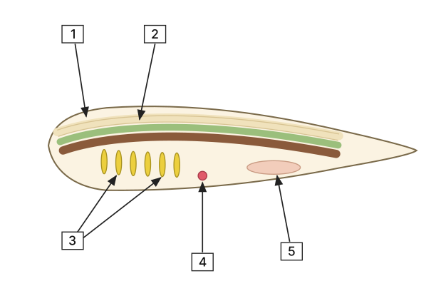
- AP EAPCET 2021Set ap-2021-09-03-FN1 markMCQQ.In the given diagram of the typical chordate. Identify the parts labelled as 1, 2, 3, 4, 5 and select the correct option? [FIGURE] (a lateral-view line diagram of a typical chordate, amphioxus-like: two elongated structures running parallel along the top of the body, pointed to by boxes labelled 1 and 2; a set of several short, closely spaced vertical slit-like markings in the middle of the body, pointed to by a box labelled 3 whose arrow fans out toward that region; a small marked point on the underside of the body just below the slit region, pointed to by a box labelled 4; and the posterior/lower body cavity region pointed to by a box labelled 5) (A) 1 - Notochord, 2 - Nerve cord, 3 - Gil slits, 4 - Heart, 5 - Gut (B) 1 - Notochord, 2 - Nerve cord, 3 - Heart, 4 - Gil slits, 5 - Anus (C) 1 - Nerve cord, 2 - Notochord, 3 - Gil slits, 4 - Heart, 5 - Gut (D) 1 - Nerve cord, 2 - Notochord, 3 - Gut, 4 - Gil slits, 5 - Heart
›Reveal solutionSolution
The diagram shows a typical chordate (like amphioxus) in lateral view; the dorsal structures are the nerve cord (outer) and notochord (inner), the vertical slits are gill slits, the ventral spot is the heart, and the posterior cavity is the gut — matching option (C).
The key to this question is knowing the basic body plan of a chordate — especially the relative positions of the major structures in a side view. In all chordates (at some stage), the nerve cord is dorsal and hollow, the notochord lies just ventral to it, pharyngeal gill slits appear laterally, and the heart is ventral. The diagram is a stylized lateral view of a generalized chordate (often based on amphioxus or a larval tunicate). Let’s map each label step by step.
-
Identify the two dorsal structures (labels 1 and 2).
In the diagram, the topmost pale band (label 1) is the outermost layer along the back. Just beneath it is a greenish band (label 2). In chordates, the nerve cord runs along the dorsal side, and the notochord lies immediately below it. The nerve cord is typically more superficial (closer to the skin) and the notochord is deeper. Therefore, label 1 = nerve cord and label 2 = notochord. This eliminates options (A) and (B), which swap these.
-
Identify the vertical slit-like markings (label 3).
A row of short, closely spaced vertical slits in the mid-body region is the classic signature of pharyngeal gill slits — a defining chordate feature. These are openings from the pharynx to the outside. So label 3 = gill slits. This rules out option (D), which calls label 3 “gut”.
-
Identify the small ventral spot (label 4).
Just below the gill slits, on the underside, there is a small red/pink spot. In chordates, the heart is located ventrally, often near the pharyngeal region. This matches the position and the typical coloring used in diagrams. So label 4 = heart.
-
Identify the posterior cavity (label 5). …
-
- AP EAPCET 2021Set ap-2021-09-06-FN1 markMCQQ.The nerve cord of chordates is _________ (A) Single, solid, tubular and non-ganglionated (B) Double, hollow, tubular and non-ganglionated (C) Single, hollow, tubular and ganglionated (D) Single, hollow, tubular and non-ganglionated
›Reveal solutionSolution
This tests one of the five basic chordate characters — the nature of the nerve cord; the correct description is (D).
Concept and Intuition
Chordata is defined partly by a dorsal, hollow, tubular nerve cord, in sharp contrast to the solid, ventral, ganglionated nerve cord seen in non-chordate invertebrates such as annelids and arthropods.
Step-by-Step Solution
- "Single" — chordates have one nerve cord, not a paired one.
- "Hollow, tubular" — it is a fluid-filled neural tube/canal, not a solid rod. …
- AP EAPCET 2021Set ap-2021-09-06-FN1 markMCQQ.Ascidia belongs to the subphylum _________ (A) Urochordata (B) Cephalochordata (C) Craniata (D) Unichordata
›Reveal solutionSolution
This tests which chordate subphylum Ascidia belongs to; the answer is (A) Urochordata.
Concept and Intuition
Chordata is split into three subphyla based on where and when the notochord is present: Urochordata (notochord confined to the larval tail; adults, like Ascidia, are sessile filter feeders), Cephalochordata (notochord persists life-long, e.g. Amphioxus), and Vertebrata/Craniata (notochord replaced by a vertebral column).
Step-by-Step Solution
- Ascidia is a classic sea squirt — a tunicate. …
🎓Unlock everything free for 14 days
- ✓Full step-by-step solutions
- ✓Concept-first explanations
- ✓Methods, shortcuts & mistakes
- ✓PYQ mapping + timed mock tests
Full access for 14 days. No credit card required.