Q.Name the only hormone secreted by pars intermedia of the pituitary gland.
🔒You're viewing a preview — the full solution, concept, methods & PYQ mapping are locked.
🔒 Start your 14-day free trial to unlock the full solution →Concept understanding — Pituitary Gland Hormones
The Master Gland and Its Messengers
The pituitary gland sits at the base of your brain, about the size of a pea. But don't let its size fool you — it's called the "master gland" because it controls so many other glands in your body. Think of it as the CEO of a company: it doesn't do all the work itself, but it sends out orders that make other departments (thyroid, adrenal glands, ovaries, testes) do their jobs.
The pituitary has two distinct parts: the anterior lobe (front) and the posterior lobe (back). They're like two different departments that happen to share the same building. The anterior lobe makes its own hormones; the posterior lobe stores and releases hormones that were actually made in the brain (the hypothalamus).
Anterior Pituitary Hormones (Made Here)
The anterior pituitary produces six major hormones. Each one targets a specific organ or tissue elsewhere in the body.
Growth Hormone (GH)
GH stimulates growth in almost every tissue — especially bones and muscles. It's most active during childhood and adolescence. If too little is secreted, a child may have stunted growth (pituitary dwarfism). Too much can lead to gigantism in children or acromegaly (enlarged hands, feet, jaw) in adults.
Thyroid-Stimulating Hormone (TSH)
TSH travels to the thyroid gland in your neck and tells it to release thyroid hormones (T3 and T4). Those thyroid hormones then regulate your metabolism — how fast your body burns energy. Without TSH, your thyroid goes dormant.
Adrenocorticotropic Hormone (ACTH)
ACTH targets the adrenal cortex (the outer layer of the adrenal glands, which sit on top of your kidneys). It triggers the release of cortisol, a stress hormone that helps manage blood sugar, inflammation, and immune responses.
Luteinizing Hormone (LH) and Follicle-Stimulating Hormone (FSH)
These two work together as the gonadotropins — they control the reproductive system.
- In females: FSH stimulates the growth of ovarian follicles (which contain eggs); LH triggers ovulation (release of the egg) and helps form the corpus luteum.
- In males: FSH helps produce sperm; LH stimulates testosterone production from the testes.
Prolactin
Prolactin's main job is to initiate and maintain milk production in the mammary glands after childbirth. It's normally kept in check by dopamine from the hypothalamus — that's why prolactin levels stay low unless a woman is breastfeeding.
The anterior pituitary is sometimes called the adenohypophysis. Its hormones are all protein or peptide hormones — they can't cross cell membranes, so they bind to surface receptors and trigger second messengers inside the cell.
Posterior Pituitary Hormones (Stored Here, Made Upstairs)
The posterior pituitary doesn't make its own hormones. Instead, the hypothalamus (the brain region just above it) produces two hormones, which then travel down nerve fibres into the posterior pituitary for storage and release.
Oxytocin
Oxytocin is best known for two actions:
- Childbirth: It stimulates strong contractions of the uterine muscles during labour.
- Milk ejection: When a baby suckles, oxytocin is released, causing milk to be "let down" from the mammary glands.
It's also involved in bonding and social behaviour — sometimes called the "love hormone."
Vasopressin (Antidiuretic Hormone, ADH)
Vasopressin acts on the kidneys. It tells them to reabsorb more water back into the blood, producing more concentrated urine. This is how your body conserves water when you're dehydrated.
If vasopressin is deficient, you get diabetes insipidus — not the sugar-related diabetes, but a condition where you produce huge volumes of dilute urine and feel constantly thirsty.
Don't confuse diabetes insipidus (ADH deficiency) with diabetes mellitus (insulin deficiency). Both cause frequent urination, but the underlying hormones and mechanisms are completely different.
Summary Table
| Part | Hormone | Target | Main Effect |
|---|---|---|---|
| Anterior | GH | Bones, muscles, tissues | Growth |
The only hormone secreted by the pars intermedia is melanocyte stimulating hormone (MSH).
- The pars intermedia is a part of the adenohypophysis, though in humans it is almost fused with the pars distalis (anterior pituitary).
- It secretes just this one hormone. …
The pars intermedia of the pituitary secretes just one hormone, melanocyte stimulating hormone, which regulates skin pigmentation.
The pituitary gland's adenohypophysis is made of two portions: the pars distalis, commonly called the anterior pituitary, and the pars intermedia. While the pars distalis is a busy hormone factory producing six different hormones, the pars intermedia is comparatively simple in output, secreting only a single hormone, melanocyte stimulating hormone (MSH). …
- AP EAPCET 2026Set ap-2026-05-19-FN1 markMCQQ.Which hormone is released by posterior lobe of pituitary? (A) Growth hormone (B) Prolactin (C) Follicle stimulating hormone (D) Oxytocin
›Reveal solutionSolution
Posterior pituitary releases oxytocin (and ADH); the other three listed hormones
are anterior pituitary hormones. Answer: (D).
Concept and Intuition
The pituitary gland has two distinct embryological and functional parts:
- Anterior pituitary (adenohypophysis): makes and releases GH, Prolactin, TSH, ACTH, FSH, LH.
- Posterior pituitary (neurohypophysis): does not synthesize hormones itself; it stores and releases oxytocin and vasopressin (ADH), both made in the hypothalamus and transported down the hypothalamo-hypophyseal tract.
Step-by-Step Solution
- Growth hormone (A) — anterior pituitary, somatotrophs.
- Prolactin (B) — anterior pituitary, lactotrophs. …
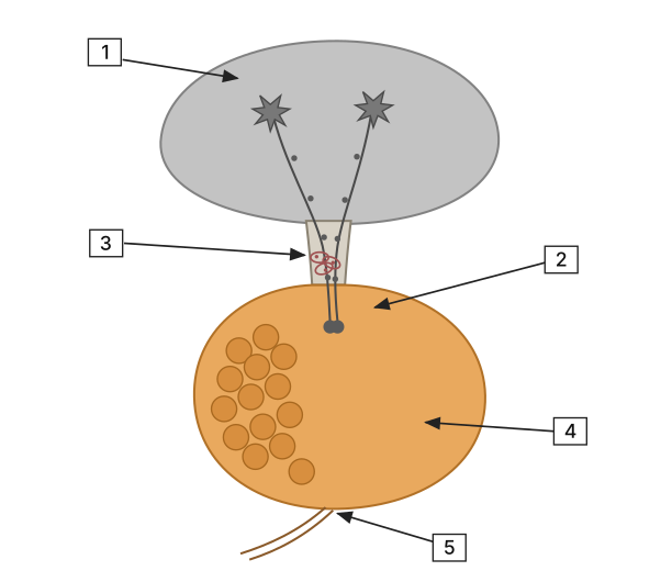
- AP EAPCET 2021Set ap-2021-09-03-FN1 markMCQQ.Pituitary gland and Hypothalamus are shown in the figure with labels 1 to 5. Identify them correctly? [FIGURE] (a diagram of the pituitary gland and hypothalamus: an upper grey shaded region containing neuron-like cell bodies with long descending fibres, pointed to by a box labelled 1; below it, a narrow stalk-like connecting region pointed to by a box labelled 3; a network of small vessel-like loops within the stalk area pointed to by a box labelled 2; a larger lower-right lobe pointed to by a box labelled 4; and a lower-left lobe containing several rounded follicle-like structures pointed to by a box labelled 5) (A) 1 - Infundibulum, 2 - Hypothalamus, 3 - Portal Circulation, 4 - Posterior Pituitary, 5 - Anterior Pituitary (B) 1 - Hypothalamus, 2 - Infundibulum, 3 - Portal Circulation, 4 - Anterior Pituitary, 5 - Posterior Pituitary (C) 1 - Hypothalamus, 2 - Infundibulum, 3 - Portal Circulation, 4 - Posterior Pituitary, 5 - Anterior Pituitary (D) 1 - Infundibulum, 2 - Hypothalamus, 3 - Portal Circulation, 4 - Anterior Pituitary, 5 - Posterior Pituitary
›Reveal solutionSolution
The key is to match the anatomical labels to the correct structures: the grey upper region is the Hypothalamus (1), the stalk is the Infundibulum (2), the capillary network in the stalk is the Portal Circulation (3), the solid right lobe is the Posterior Pituitary (4), and the left lobe with follicle-like structures is the Anterior Pituitary (5). The correct option is (C).
The question tests your understanding of the hypothalamic-pituitary axis — the master control system of the endocrine system. The hypothalamus sits above the pituitary, connected by a stalk (the infundibulum). The pituitary has two distinct lobes: the posterior pituitary (neural, made of axon terminals from hypothalamic neurons) and the anterior pituitary (glandular, with hormone-secreting cells). The blood supply between them is special: a portal circulation carries releasing hormones from the hypothalamus directly to the anterior pituitary.
The figure’s labels correspond to these structures. Let’s decode them step by step.
-
Label 1 points to the grey upper region with neuron-like cell bodies.
This is clearly the Hypothalamus — the brain region that produces hormones and sends axons down the stalk. The long descending fibers are hypothalamic neurons. So label 1 = Hypothalamus.
-
Label 2 points to the narrow stalk-like connecting region.
This is the Infundibulum — the anatomical stalk that physically connects the hypothalamus to the pituitary. It contains the hypothalamic axons going to the posterior pituitary and the portal blood vessels. So label 2 = Infundibulum.
-
Label 3 points to the network of small vessel-like loops within the stalk area.
This is the Hypothalamic-pituitary portal circulation — a capillary network that carries hypothalamic releasing hormones to the anterior pituitary. It’s not a solid structure but a vascular system. So label 3 = Portal Circulation.
-
Label 4 points to the larger lower-right lobe (solid, without follicles).
This is the Posterior Pituitary (neurohypophysis). It is composed of axon terminals from hypothalamic neurons (the long fibers from label 1 end here). It stores and releases oxytocin and ADH. It appears solid because it’s neural tissue. So label 4 = Posterior Pituitary.
-
Label 5 points to the lower-left lobe with rounded follicle-like structures. …
-
- AP EAPCET 2021Set ap-2021-09-06-AN1 markMCQQ.Assertion (A): Adrenocorticotropic - Releasing Hormone (ARH) excites the anterior lobe of pituitary gland to produce Adrenocorticotropic Hormone (ACTH) Reason (R): ACTH stimulates the adrenal cortex to secrete its glucocorticoid and mineralocorticoid hormones (A) Both A and R are true but the R is not a correct explanation of the A (B) A is true but R is false (C) Both A and R are false (D) A is false but R is true
›Reveal solutionSolution
This tests the hypothalamus–pituitary–adrenal axis; both statements are factually true, but the Reason (about ACTH's action on the adrenal cortex) does not explain the Assertion (about the releasing hormone's action on the pituitary).
Concept and Intuition
The hypothalamic-pituitary-adrenal (HPA) axis is a sequential hormonal cascade. A hypothalamic releasing hormone travels via the hypophyseal portal system to the anterior pituitary, where it stimulates specialised cells to secrete adrenocorticotropic hormone (ACTH). ACTH then circulates to the adrenal cortex, where it stimulates the synthesis and secretion of corticosteroids, encompassing both glucocorticoids (e.g., cortisol, involved in metabolism and stress response) and, to some extent, mineralocorticoids (e.g., aldosterone, involved in electrolyte/water balance). Both of these are accurate, standard descriptions of two separate, sequential links in this axis: (1) releasing hormone → ACTH, and (2) ACTH → adrenal cortex steroids. Because the Reason only describes link (2), it cannot serve as the mechanistic explanation for link (1) described in the Assertion — they describe two different steps of the same cascade.
Step-by-Step Solution
- Evaluate Assertion: hypothalamic releasing hormone stimulating the anterior pituitary to secrete ACTH — a correct description of the first HPA axis link.
- Evaluate Reason: ACTH stimulating the adrenal cortex to secrete glucocorticoids and mineralocorticoids — also a correct description, but of the second, later link in the axis. …
🎓Unlock everything free for 14 days
- ✓Full step-by-step solutions
- ✓Concept-first explanations
- ✓Methods, shortcuts & mistakes
- ✓PYQ mapping + timed mock tests
Full access for 14 days. No credit card required.