From the Outside In: Why Leaves Look Different
When you pick up a leaf from a mango tree and one from a grass blade, the first thing you notice is the shape. The mango leaf is broad and flat; the grass blade is long, narrow, and stands upright. That difference isn't just cosmetic — it reflects two fundamentally different ways of building a leaf, each suited to a different environment.
A leaf's job is photosynthesis. To do it well, it needs to capture sunlight, exchange gases, and manage water loss. The internal architecture of a dicot leaf and a monocot leaf are two different solutions to these same problems.
Dicot Leaves: The Dorsiventral Design
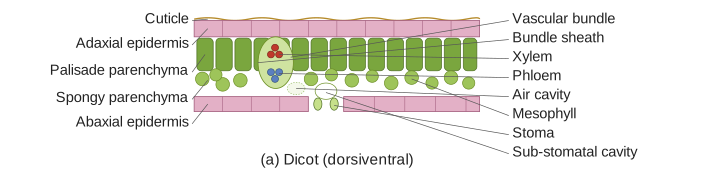
Most dicot leaves (like those of sunflower, mango, or hibiscus) are dorsiventral — they have a distinct top (dorsal) and bottom (ventral) surface. Hold a leaf up: the upper side is darker green, shinier, and faces the sun. The lower side is paler and has more stomata.
Why? Because the upper surface is the light-catcher. The cells that do most of the photosynthesis are packed right beneath it. The lower surface is the gas-exchange zone — stomata open into air spaces, letting CO₂ in and O₂ out, while minimising water loss from the sun-baked top.
Mesophyll Differentiation
The internal tissue between the upper and lower epidermis — the mesophyll — is clearly divided into two layers:
- Palisade parenchyma: Tall, column-shaped cells packed tightly just below the upper epidermis. They contain most of the chloroplasts. This is the factory floor — light hits here first, and the cells are arranged to absorb as much of it as possible.
- Spongy parenchyma: Irregular, loosely packed cells with large air spaces between them, near the lower epidermis. Fewer chloroplasts. The air spaces allow CO₂ to diffuse from the stomata up to the palisade cells.
This two-layer system is the hallmark of a dorsiventral leaf. The palisade layer is usually one or two cells thick; the spongy layer is several cells thick.
In a dorsiventral leaf, the xylem (water-conducting tissue) of each vein faces the upper epidermis, and the phloem (food-conducting tissue) faces the lower epidermis. This orientation is consistent across all veins.
Monocot Leaves: The Isobilateral Design
Monocot leaves (like those of maize, wheat, or grass) are isobilateral — both surfaces look more or less the same. There is no clear upper and lower side. The leaf stands upright, so both surfaces receive roughly equal light.
Mesophyll: No Palisade-Spongy Split
Here is the key difference: monocot mesophyll is not differentiated into palisade and spongy layers. Instead, the mesophyll cells are all more or less alike — irregular, with chloroplasts distributed throughout. They are packed together with small air spaces.
Why no differentiation? Because the leaf is vertical. Light doesn't hit one surface preferentially. Both sides need to photosynthesise equally, so there is no need for a specialised light-capturing layer on one side only.
Bulliform Cells: The Leaf's Hinge
Monocot leaves have a special adaptation you won't find in dicots: bulliform cells. These are large, thin-walled, colourless cells (no chloroplasts) found in groups on the upper epidermis, often in the grooves between veins.
When water is plentiful, bulliform cells become turgid (swollen with water). This pushes the leaf surface flat, maximising light capture. When water is scarce, they lose turgor and become flaccid. The leaf then folds inward (like a book closing), reducing the surface area exposed to the sun and cutting water loss.
Think of bulliform cells as a built-in water-saving mechanism. When the grass is well-watered, the leaf blade is flat. During drought, the leaf rolls up — those bulliform cells are the hinge that makes it happen.
Side-by-Side Comparison
| Feature | Dicot Leaf (Dorsiventral) | Monocot Leaf (Isobilateral) |
|---|---|---| …