Biology · Ch 8 — Cell: The Unit of Life
Nucleus
Nucleus
The nucleus is the control centre of the eukaryotic cell — it directs the activities of the other organelles and carries the hereditary material. It was first described as a cell organelle by Robert Brown, as early as 1831.
Naming of chromatin
The nuclear material that takes up basic (staining) dyes was later given the name chromatin by Flemming. In a cell that is not dividing — the interphase nucleus — this chromatin exists as highly extended, elaborate nucleoprotein fibres. Along with these fibres, the interphase nucleus contains a nuclear matrix and one or more spherical bodies called nucleoli (singular: nucleolus).
The nuclear envelope
Electron microscopy reveals that the nucleus is bounded by a nuclear envelope made of two parallel membranes. Between these two membranes lies a gap called the perinuclear space, roughly ten to fifty nanometres wide. This double-membrane envelope acts as a barrier separating the contents of the nucleus from the cytoplasm.
Key features of the envelope include:
- The outer membrane is usually continuous with the endoplasmic reticulum and also bears ribosomes on its surface.
- At many points the envelope is interrupted by minute nuclear pores, each formed where the two membranes fuse together.
- These pores act as passageways through which RNA and protein molecules move in both directions between the nucleus and the cytoplasm.
Number of nuclei
Normally there is just one nucleus per cell, but variations occur — some cells have more than one nucleus. A few mature cells even lack a nucleus altogether; familiar examples are the erythrocytes (red blood cells) of many mammals and the sieve tube cells of the vascular tissue of plants.
Nucleoplasm and nucleolus
The nuclear matrix, also called the nucleoplasm, contains both the nucleolus and the chromatin. The nucleoli are spherical structures suspended in the nucleoplasm. A nucleolus is not a membrane-bound structure, so its contents are continuous with the surrounding nucleoplasm. Its main job is to serve as a site of active ribosomal RNA (rRNA) synthesis. Cells that are very active in protein synthesis tend to have larger and more numerous nucleoli, reflecting their greater need to make ribosomes.
Chromatin and chromosomes
The loose, indistinct network of nucleoprotein fibres seen in the interphase nucleus is the chromatin. During cell division, however, this material condenses so that well-defined chromosomes appear in place of the diffuse network.
Chromatin is made up of:
- DNA
- Histones (basic proteins)
- Non-histone proteins
- RNA
A single human cell contains about two metres of DNA thread in total, distributed among its forty-six chromosomes (that is, twenty-three pairs). The detailed way DNA is packaged into a chromosome is studied in a later class.
Parts of a chromosome
Every chromosome — visible only in dividing cells — has certain characteristic parts:
- A primary constriction, also called the centromere.
- On either side of the centromere are disc-shaped structures called kinetochores.
- The centromere is the region that holds the two chromatids of a chromosome together.
Types of chromosomes based on centromere position
Depending on where the centromere sits, chromosomes are grouped into four types: …
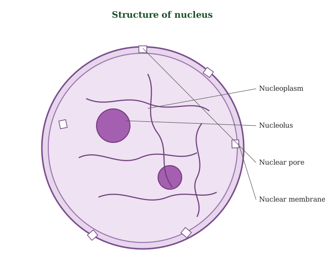
Drawn by us to help you understand the concept clearly, and verified to make sure it's accurate. For exams, practice from your NCERT textbook's own diagram.
This figure shows the nucleus, the control centre of the cell. It is enclosed by a double-membrane nuclear envelope, and this envelope is pierced by many nuclear pores through which materials pass between the nucleus and the cytoplasm. Inside lies a fluid called the nucleoplasm. Within it sit one or more rounded, dense bodies called nucleoli, where ribosomal RNA is made and ribosome parts begin to form. Also spread through the nucleoplasm is a fine, thread-like network called chromatin, made of DNA and protein. When the cell prepares to divide, this loose chromatin coils and condenses into distinct rod-like chromosomes. The idea to hold on to is t …
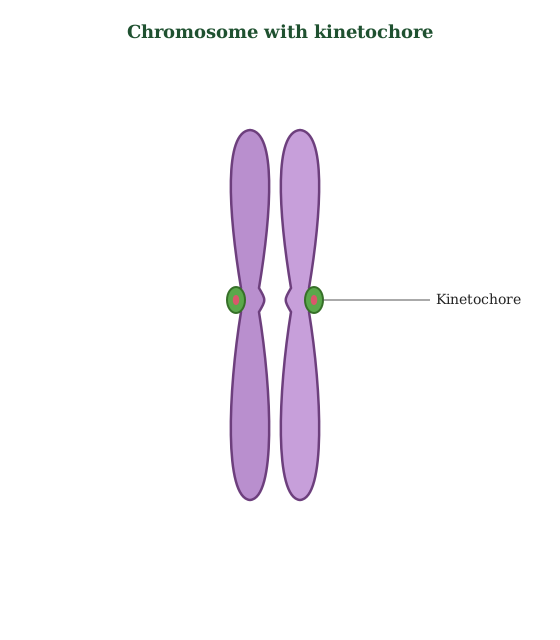
Drawn by us to help you understand the concept clearly, and verified to make sure it's accurate. For exams, practice from your NCERT textbook's own diagram.
This figure shows the structure of a chromosome as it appears when a cell is about to divide. The chromosome is made of two identical threads called sister chromatids, which are copies of each other joined together. They are held at a single narrowed region called the centromere, seen as a primary constriction where the chromatids pinch inward. On the surface of the centromere lie two small disc-shaped plates, one on each side, called kinetochores. These are the points where fibres of the spindle attach during cell division so the chromosome can be pulled apart correctly. The key idea is that the centromere and its ki …
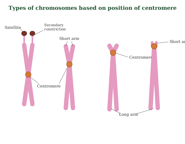
Drawn by us to help you understand the concept clearly, and verified to make sure it's accurate. For exams, practice from your NCERT textbook's own diagram.
This figure sorts chromosomes into four types according to where the centromere sits, because its position decides how long the two arms are. In a metacentric chromosome the centromere lies in the middle, so the two arms are roughly equal and it looks like the letter V during movement. In a sub-metacentric chromosome the centromere is shifted slightly to one side, giving one arm a little shorter than the other and an L-like shape. In an acrocentric chromosome the centromere lies close to one end, leaving one very long arm and one very short arm. In a telocentric chromosome the centromere sits right at the tip, so effectiv …