Q.Draw a labelled ray diagram showing the formation of image in a compound microscope. Define its magnifying power and write expression for it. OR Using Huygen's construction, explain reflection of a plane wave by a plane surface.
You're viewing a preview — the full solution, concept, methods & PYQ mapping are locked.
Start your 14-day free trial to unlock the full solution →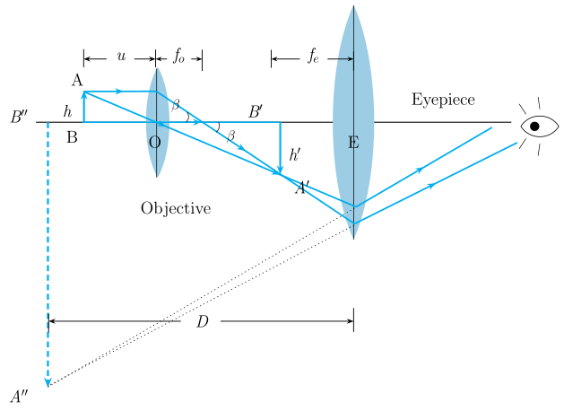
In a compound microscope, the objective forms a real magnified image, which the eyepiece magnifies further as a simple magnifier; overall magnifying power .
Ray diagram (described): The object is placed just outside the focus of the objective lens (a short focal-length convex lens). The objective forms a real, inverted, and magnified image inside the microscope tube, beyond its own focal length but within the focal length of the eyepiece. This intermediate image acts as the object for the eyepiece (a convex lens used as a simple magnifier), which forms a final image — virtual, further magnified, and inverted relative to the original object — typically at the near point (least distance of distinct vision) , or at infinity for relaxed-eye viewing. Rays from the top of the object pass through the objective converging to the top of , then diverge from through the eyepiece to form the enlarged virtual image .
Magnifying power: Defined as the ratio of the angle subtended at the eye by the final image to the angle subtended by the object when placed at the near point (viewed directly, without the instrument).
The overall magnifying power is the product of the objective's linear magnification and the eyepiece's angular magnification (acting as a simple magnifier):
For the final image formed at the near point :
…
Unlock everything free for 14 days
- Full step-by-step solutions
- Concept-first explanations
- Methods, shortcuts & mistakes
- PYQ mapping + timed mock tests
Full access for 14 days. No credit card required.