Q.Draw a well labelled diagram of microscopic anatomy of testis.
🔒You're viewing a preview — the full solution, concept, methods & PYQ mapping are locked.
🔒 Start your 14-day free trial to unlock the full solution →Concept understanding — Male Reproductive Anatomy
Male Reproductive Anatomy – A First Look
Think of the male reproductive system as a production and delivery line. Its job is to make tiny cells called sperm (the male contribution to reproduction) and then safely deliver them into the female body. Unlike the female system, which is mostly internal, the male system has several parts that are visible outside the body. That’s the first big clue: the system is built for external production and internal delivery.
The Two Main Jobs
The male reproductive system does two things at once:
- Spermatogenesis – making sperm inside the testes.
- Hormone production – making the male sex hormone testosterone, which drives puberty, muscle growth, voice deepening, and sex drive.
These two jobs happen in the same organs, but they are separate processes. Testosterone is made by special cells between the sperm-making tubes, not inside the tubes themselves.
The Key Parts (from outside to inside)
1. The Testes (the factories)
The two testes (singular: testis) are oval organs about the size of a large olive. They hang outside the body in a pouch of skin called the scrotum. Why outside? Because sperm production needs a temperature about 2–3°C lower than normal body temperature. The scrotum can contract (pull testes closer to the body when cold) or relax (let them hang lower when warm) to regulate this temperature.
Inside each testis are about 250–300 coiled tubes called seminiferous tubules. This is where sperm are actually made. Between these tubules are the interstitial cells (Leydig cells) that produce testosterone.
The testes are the primary male reproductive organs – they produce both the gametes (sperm) and the hormone (testosterone). Everything else in the system is a duct, gland, or delivery tube.
2. The Duct System (the conveyor belt)
Once sperm are made, they need to travel. The path is:
Seminiferous tubules → Rete testis → Vasa efferentia → Epididymis → Vas deferens → Ejaculatory duct → Urethra
The epididymis is a long, coiled tube stuck to the back of each testis. Sperm are stored here and become motile (able to swim) as they pass through. It takes about 2–3 weeks for a sperm to travel the full length of the epididymis.
The vas deferens (plural: vasa deferentia) is a thick-walled muscular tube that runs from the scrotum up into the abdomen. You can feel it as a firm cord just above the testis – it’s part of the spermatic cord. During a vasectomy, this tube is cut or blocked to prevent sperm from reaching the urethra.
3. The Accessory Glands (the fluids)
Sperm alone are just a tiny fraction of the fluid that comes out during ejaculation. Most of the volume comes from three sets of glands:
- Seminal vesicles – two sac-like structures behind the bladder. They secrete a thick, fructose-rich fluid that provides energy for the sperm. This makes up about 60–70% of the semen volume.
- Prostate gland – a walnut-sized gland that surrounds the urethra just below the bladder. It secretes a thin, milky fluid that makes the semen more fluid and slightly alkaline (to protect sperm from the acidic vagina).
- Bulbourethral glands (Cowper’s glands) – two pea-sized glands near the base of the penis. They secrete a clear, slippery fluid before ejaculation that lubricates the urethra and neutralises any leftover urine acidity.
Semen is not just sperm. It is a mixture of sperm (about 1–5% of the volume) plus secretions from the seminal vesicles, prostate, and bulbourethral glands. The sperm themselves are made in the testes; everything else is added along the way.
4. The Penis (the delivery tube)
The penis is the external organ that deposits semen into the female vagina. It contains three columns of erectile tissue that fill with blood during arousal, causing an erection. The urethra runs through the centre of the penis and carries both urine and semen – but never at the same time. A sphincter (a ring of muscle) at the base of the bladder closes off the urine passage during ejaculation.
The tip of the penis is called the glans penis, which is covered by a loose fold of skin called the foreskin (prepuce) in uncircumcised males.
Why This Matters (beyond reproduction) …
The testis carries out two structurally separate jobs — producing sperm inside its coiled tubules and secreting hormones from cells lying in the spaces between them — and a correctly labelled diagram must show both regions along with every stage of sperm maturation within the tubule wall. …
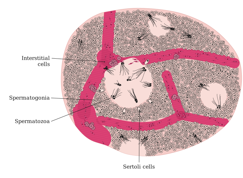
A microscopic (T.S.) section of the testis shows seminiferous tubules containing the spermatogenic (germinal) epithelium and Sertoli cells, surrounded by interstitial (Leydig) cells in the stroma between tubules.
Note on the diagram: a hand-drawn figure cannot be produced in this text answer, so here is the complete structure with every label a well-drawn diagram must carry, in the correct spatial arrangement, so you can reproduce it accurately.
Overall organisation:
- The testis is covered externally by a dense fibrous capsule, the tunica albuginea.
- Septa extending inward from the capsule divide each testis into about 250 testicular lobules, each containing one to three highly coiled seminiferous tubules.
Microscopic (T.S. of a seminiferous tubule) — label these parts from outside to inside:
- Tunica propria (basement membrane) — thin outer covering of the tubule.
- Germinal (spermatogenic) epithelium lining the inside of the tubule wall, arranged in layers from the base towards the lumen:
- Spermatogonia (2n) — outermost/basal layer, undifferentiated diploid germ cells that multiply mitotically.
- Primary spermatocytes (2n) — formed from spermatogonia, undergo meiosis-I.
- Secondary spermatocytes (n) — haploid, formed after meiosis-I, undergo meiosis-II.
- Spermatids (n) — haploid, formed after meiosis-II, lie closer to the lumen.
- Spermatozoa (sperm) — mature, released into the lumen of the tubule after spermiogenesis. …
Showing the 12 most recent of 39 on this concept.
- CBSE 2026Set V11 markMCQQ.The cells that secrete androgens are(a) Sertoli cells(b) Leydig cells(c) Germ cells(d) Spermatogonia
›Reveal solutionSolution
The interstitial Leydig cells of the testis secrete androgens.
In the testis, the Leydig cells (interstitial cells) lie in the spaces between the seminiferous tubules and synthesise and secrete the male sex hormones called androgens (mainly testosterone). Sertoli cells nourish the de …
- CBSE 2026Set A1 markMCQQ.Which of the following cells are found in seminiferous tubules?(a) Sertoli cells(b) Interstitial cells(c) Spermatogonia(d) Both (A) and (C)
›Reveal solutionSolution
Inside the seminiferous tubules are spermatogonia (germ cells) and Sertoli (nurse) cells; interstitial (Leydig) cells lie outside the tubules.
The lining of each seminiferous tubule is made of two types of cells: the male germ cells or spermatogonia, which undergo meiosis to form sperm, and the Sertoli cells (nurse cells), which provide nourishment to the developing germ cells. The interstitial cells (Leydig cells), which secrete testosterone, are lo …
- CBSE 2026Set A1 markMCQQ.In which structure of sperm are the enzymes present which help in fertilisation of the ovum?(a) Middle piece(b) Neck(c) Nucleus(d) Acrosome
›Reveal solutionSolution
The acrosome, a cap over the sperm head, holds enzymes that help the sperm enter the egg.
The head of a sperm is covered by a cap-like structure called the acrosome. It is filled with hydrolytic enzymes (such as hyaluronidase) that digest and break down the outer coverings of the ovum (the zona pellucida), allowing the sperm to penetrate and fertilise the egg. The middle piece contains mit …
- CBSE 2026Set ANNUAL1 markMCQQ.Acrosome is found in which part of the human sperm?(a) Tail(b) Middle piece(c) Head(d) Neck
›Reveal solutionSolution
The acrosome is an enzyme-filled cap over the sperm's head that helps the sperm penetrate the egg's outer coverings.
A human sperm has a head, neck, middle piece and tail. The head contains an elongated, haploid nucleus, the anterior portion of which is covered by a cap-like structure called the acrosome, filled with enzymes (such as hyaluronidase) that facilitate fertilisation by digesting t …
- CBSE 2026Set ANNUAL1 markMCQQ.Which of the following is not a part of male reproductive system?(a) Ampulla(b) Prostate(c) Foreskin(d) Bulbourethral gland
›Reveal solutionSolution
Prostate, bulbourethral (Cowper's) glands and the foreskin are all male reproductive structures; the ampulla belongs to the female oviduct.
In the male reproductive system, the accessory glands (prostate, seminal vesicles, bulbourethral glands) add secretions to semen, and the foreskin (prepuce) covers the glans penis. The ampulla, by contrast, is the wide distal part of the female fall …
- CBSE 2026Set ANNUAL1 markMCQQ.Leydig cells are found :(a) in liver(b) in testes(c) in kidney(d) in uterus
›Reveal solutionSolution
Leydig cells (interstitial cells) are found in the testes, between the seminiferous tubules.
Inside the testis, in the interstitial spaces between the seminiferous tubules, are small clusters of glandular cells called Leydig cells (interstitial cells). They are steroidogenic cells that synthesise and secrete the male sex hormone testosterone (androgen), which controls the development of secondary sexual characters in males …
- CBSE 2026Set ANNUAL1 markQ.In which part of the body is the male reproductive system located?
›Reveal solutionSolution
The human male reproductive system is located in the pelvic region.
Concept: The male reproductive system comprises the testes, accessory ducts (rete testis, vasa efferentia, epididymis, vas deferens), accessory glands and the external genitalia. …
- CBSE 2026Set ANNUAL1 markMCQQ.The testes in humans are located outside the abdominal cavity within a pouch called the scrotum. This positioning helps in :(a) avoiding compression by internal organs.(b) maintaining a temperature slightly lower than the body temperature for sperm formation.(c) protecting the testes from physical injury during movement.(d) helping in the storage of urine before excretion.
›Reveal solutionSolution
The scrotum keeps the testes cooler than the body core, and this lower temperature is required for sperm production, so (b) is correct.
In the CBSE/NCERT Human Reproduction chapter, the testes lie outside the abdomen in the scrotum. Spermatogenesis (sperm formation) proceeds normally only at a temperature 2-2.5°C below core body temperature. The scrotal sac provides exactly this cooler environment. If the testes are retained inside the abdomen (cryptorchidism), the higher temperature impairs sperm formatio …
- CBSE 2025Set 57/4/11 markMCQQ.Given below are a few statements with reference to the accessory ducts of the human male reproductive system :(i) The seminiferous tubules of the testes open into rete testis then into the vas deferens.(ii) The vasa efferentia leave the testes and open into the epididymis.(iii) The epididymis leads to vas deferens that ascends into the abdomen.(iv) The vas deferens receives a duct from the prostrate gland and opens into the urethra as ejaculatory duct.(v) The urethra originates from the urinary bladder and extends through the penis to its external opening, urethral meatus. Choose the option with all true statements from the given options : (A) (i), (ii),(iv) (B) (ii), (iii),(v) (C) (ii), (iv),(v) (D) (i), (iii), (iv)
›Reveal solutionSolution
This question tests knowledge of the correct sequence and connections of the accessory ducts in the human male reproductive system. Statements (ii), (iii), and (v) accurately describe these ducts, making option (B) the correct choice.
Concept and Intuition
The male reproductive system includes a network of accessory ducts that play a crucial role in the transport, storage, and maturation of sperm. Understanding the precise pathway sperm take from their production site in the testes to their exit from the body is key. This pathway involves a series of interconnected tubes, each with a specific function and anatomical location. Any deviation from this correct sequence or connection makes a statement false.
-
Evaluate Statement (i): "The seminiferous tubules of the testes open into rete testis then into the vas deferens."
- Sperm are produced in the seminiferous tubules within the testes. From there, they move into the rete testis, a network of tubules. This part of the statement is correct.
- However, from the rete testis, sperm do not directly enter the vas deferens. Instead, they pass through several small ducts called the vasa efferentia, which then lead into the epididymis. Only after passing through the epididymis does the duct continue as the vas deferens.
- Therefore, this statement is false because it omits the vasa efferentia and epididymis in the sequence.
-
Evaluate Statement (ii): "The vasa efferentia leave the testes and open into the epididymis."
- Following the rete testis, the vasa efferentia (also called efferent ductules) are indeed the ducts that emerge from the testes and transport sperm to the epididymis. The epididymis is a highly coiled tube located on the posterior surface of each testis, where sperm mature and are stored.
- This statement accurately describes the anatomical connection.
- Therefore, this statement is true.
-
Evaluate Statement (iii): "The epididymis leads to vas deferens that ascends into the abdomen."
- The tail of the epididymis is continuous with the vas deferens (also known as the ductus deferens). The vas deferens is a muscular tube that ascends from the scrotum into the abdominal cavity, looping over the urinary bladder. It is a major component of the spermatic cord.
- This statement correctly describes the continuation of the epididymis and the path of the vas deferens.
- Therefore, this statement is true.
-
Evaluate Statement (iv): "The vas deferens receives a duct from the prostrate gland and opens into the urethra as ejaculatory duct." …
-
- CBSE 2025Set ANNUAL1 markMCQQ.Sertoli cells are present in :(a) Kidney(b) Testis(c) Liver(d) Ovary
›Reveal solutionSolution
Sertoli cells occur in the seminiferous tubules of the testis.
Sertoli cells (nurse cells) line the walls of the seminiferous tubules inside the testis. They provide nutrition to the developing germ cells during spermatogenesis, secrete the hormone inhibin (which regulates FSH secretion through negative feedback), …
- CBSE 2025Set F1 markMCQQ.Which of the following human cells is motile?(a) Nerve cell(b) Cutaneous cell(c) Egg cell(d) Sperm cell
›Reveal solutionSolution
The sperm is the motile human cell.
The human sperm (spermatozoon) has a head, middle piece and a long tail (flagellum) whose lashing movements propel it through the female reproductive tract toward the egg, so it is a motile cell. Nerve cells, skin (cutaneous) cells and the ovum (egg) are non- …
- CBSE 2025Set F1 markMCQQ.From which of the following do developing sperms get nutrition?(a) Sertoli cells(b) Germinal epithilium(c) Blood cell(d) Leydig cell
›Reveal solutionSolution
Developing sperms are nourished by Sertoli (nurse) cells inside the seminiferous tubules.
Inside the seminiferous tubules of the testis there are two important cell types:
- Sertoli cells (nurse cells) — attached to the germinal epithelium; they provide nutrition and support to the developing germ cells/spermatids during spermatogenesis. …
🎓Unlock everything free for 14 days
- ✓Full step-by-step solutions
- ✓Concept-first explanations
- ✓Methods, shortcuts & mistakes
- ✓PYQ mapping + timed mock tests
Full access for 14 days. No credit card required.