Q.Explain the terms primary and secondary structure of proteins. What is the difference between α-helix and β-pleated sheet structure of proteins?
🔒You're viewing a preview — the full solution, concept, methods & PYQ mapping are locked.
🔒 Start your 14-day free trial to unlock the full solution →Concept understanding — Protein Structure Levels
Protein Structure Levels: From a String to a Working Machine
Imagine you have a long string of beads. Each bead is a different colour, and the order of colours is fixed. If you just lay that string on a table, it's a floppy, useless line. But if you could somehow make that string fold itself into a tiny, precise 3D shape — say, a key that fits a specific lock — you'd have something that actually does a job. That's exactly what a protein is.
A protein starts as a long chain of smaller units called amino acids. There are 20 different kinds, each with a unique side chain (the "colour" of the bead). The exact sequence of these amino acids is determined by your DNA. But a protein isn't just a chain — it's a chain that folds into a specific shape, and that shape determines what the protein does. If the shape is wrong, the protein can't work.
The folding happens in stages, and we call these stages the four levels of protein structure.
Level 1: Primary Structure — The Sequence
This is the simplest level: just the linear order of amino acids in the chain, linked by peptide bonds. Think of it as the sentence written in the language of proteins.
Primary structure = the sequence of amino acids from the N-terminus (start) to the C-terminus (end).
Why does this matter? Because the sequence determines everything else. Change one amino acid in a critical spot, and the entire protein can misfold. Example: sickle cell anaemia is caused by a single amino acid swap in haemoglobin — valine replaces glutamic acid at position 6. One bead out of hundreds changes colour, and the whole protein folds wrong.
Level 2: Secondary Structure — Local Folding Patterns
The chain doesn't stay straight. Hydrogen bonds form between the backbone atoms (not the side chains) of nearby amino acids. These bonds cause the chain to twist or fold into regular, repeating patterns.
Two common patterns:
- Alpha helix (α-helix): The chain coils like a spring or a spiral staircase. Hydrogen bonds form between every 4th amino acid, holding the coil tight.
- Beta sheet (β-sheet): The chain folds back and forth like a pleated fan. Hydrogen bonds form between adjacent segments, creating a flat, sheet-like structure.
Secondary structure is stabilised entirely by hydrogen bonds between the carbonyl oxygen of one amino acid and the amide hydrogen of another — both part of the peptide backbone. Side chains stick out and don't participate.
These patterns are local — they happen in short stretches of the chain. A single protein can have multiple α-helices and β-sheets separated by loops.
Level 3: Tertiary Structure — The Global 3D Shape
Now the whole chain folds into its final, compact, three-dimensional shape. This is where the protein becomes functional. The tertiary structure is stabilised by interactions between the side chains of amino acids that may be far apart in the sequence but come close in space.
What holds it together?
- Hydrophobic interactions: Nonpolar side chains cluster together in the protein's interior, away from water.
- Hydrogen bonds: Between polar side chains.
- Ionic bonds: Between positively and negatively charged side chains.
- Disulfide bridges: Covalent bonds between the sulfur atoms of two cysteine amino acids — these are strong and lock parts of the chain together.
- Van der Waals forces: Weak attractions between closely packed atoms.
A common mistake: thinking tertiary structure is just "more secondary structure." It's not. Secondary structure is local folding; tertiary structure is the global arrangement of the entire chain, including how helices and sheets pack together.
Level 4: Quaternary Structure — Multiple Chains Working Together
Some proteins are made of more than one polypeptide chain. Each chain is a separate subunit, and the quaternary structure describes how these subunits assemble into a functional complex. …
Why this formula?
Protein Structure Levels: Understanding the "Why" Behind the Hierarchy
Protein structure is not defined by a single formula, but by a logical hierarchy of organization. Each level builds on the previous one, and the "formulas" here are really principles of molecular interaction that explain why proteins fold the way they do.
Let's break down each level and the reasoning behind its key features.
1. Primary Structure: The Sequence "Formula"
What it is: The linear sequence of amino acids linked by peptide bonds.
Key "formula":
Protein=NH2-[Amino Acid]1-[AA]2-...-[AA]n-COOH
Why this holds:
- Peptide bond formation is a condensation reaction:
-COOH+NH2-→-CO-NH-+H2O
- This bond is rigid and planar due to resonance (partial double-bond character). This restricts rotation, which directly influences higher-order folding.
- The sequence is determined by DNA (genetic code). Every change in sequence can alter the entire structure — this is why a single mutation (e.g., sickle cell anemia: Glu → Val at position 6) can cause disease.
Exam insight: The primary structure is the only level that is covalently determined. All higher levels are non-covalent interactions.
2. Secondary Structure: Local Folding Patterns
Key patterns: α-helix and β-pleated sheet.
Why these form — the hydrogen bond "formula":
The α-helix
- Hydrogen bonds form between the carbonyl oxygen (C=O) of residue n and the amide hydrogen (N-H) of residue n+4.
- Why n+4? This spacing allows the backbone to coil into a right-handed helix with exactly 3.6 amino acids per turn.
- Reasoning: The peptide bond's planar nature forces the backbone into a specific geometry. The n+4 pattern maximizes H-bonding while minimizing steric clashes.
The β-sheet
- Hydrogen bonds form between adjacent strands (either parallel or antiparallel).
- Why not n+4? The backbone is extended (pleated), so H-bonds occur between different segments, not within the same chain.
Key formula (Ramachandran plot):
Only certain backbone dihedral angles (ϕ,ψ) are allowed:
- α-helix: ϕ≈−57∘, ψ≈−47∘
- β-sheet: ϕ≈−130∘, ψ≈+130∘
Why these angles? Steric hindrance — atoms cannot overlap. The Ramachandran plot shows the only regions where no two atoms clash.
3. Tertiary Structure: The 3D Fold
Key "formula": The hydrophobic effect drives folding.
Why this holds:
- Water molecules form a cage-like structure around nonpolar (hydrophobic) side chains. This is entropically unfavorable (water loses freedom).
- To minimize this, hydrophobic side chains cluster together in the protein's core, away from water.
- Result: The protein collapses into a compact globule, with polar/charged residues on the surface.
Supporting interactions (the "glue"):
| Interaction | Why it matters |
|---|---|
| Hydrogen bonds | Between side chains (e.g., Ser–Glu) |
| Ionic bonds | Between charged groups (e.g., Lys–Asp) |
| Van der Waals forces | Close packing of atoms |
| Disulfide bridges | Covalent S–S bonds (only in oxidizing environments) |
Why not just one formula? Tertiary structure is unique to each protein — it's the sum of all these interactions, not a single equation.
4. Quaternary Structure: Multiple Subunits
Key "formula":
Functional protein=∑i=1nSubuniti
Why this holds:
- Some proteins need multiple polypeptide chains to function (e.g., hemoglobin: α2β2). …
Concept: Protein Structure Levels
Primary structure is the linear sequence of amino acids linked by peptide bonds in a polypeptide chain. This sequence is determined by genetic code and dictates all higher-order folding.
Secondary structure refers to local, regular folding patterns stabilized by hydrogen bonds between backbone amide and carbonyl groups. The two most common types are the α-helix and β-pleated sheet.
Key differences between α-helix and β-pleated sheet:
- α-helix: A right-handed coiled structure where hydrogen bonds form between the −NH of one residue and the −CO of the fourth residue ahead (i to i+4). The side chains project outward from the helix. …
Primary structure is the linear sequence of amino acids; secondary structure is the local folding into α-helices or β-sheets. The α-helix is a right-handed coil stabilized by intra-chain H-bonds, while the β-sheet is a pleated arrangement of adjacent strands held by inter-strand H-bonds.
Let’s start with the big picture. Proteins are the workhorses of biology, and their function depends entirely on their shape. That shape is built in layers, from the simplest sequence to the final 3D form. The first two layers — primary and secondary structure — are the foundation.
Primary structure is the simplest level: it’s just the linear sequence of amino acids linked by peptide bonds. Think of it as a string of beads, where each bead is one of 20 different amino acids. The order is determined by the gene that codes for the protein. This sequence is written from the N-terminus (free amino group) to the C-terminus (free carboxyl group). No folding, no twisting — just the chain.
Secondary structure is where the chain starts to fold into regular, repeating patterns. These patterns are stabilized by hydrogen bonds between the backbone atoms (the −NH and −CO groups of the peptide bonds), not the side chains. The two most common patterns are the α-helix and the β-pleated sheet.
Now, the key difference between them lies in how the hydrogen bonds are arranged and how the chain is oriented.
-
The α-helix is a right-handed coil. Imagine a spiral staircase. The backbone winds around a central axis, and the side chains stick outward. Every peptide bond’s −NH group forms a hydrogen bond with the −CO group of the amino acid four residues earlier in the chain. This creates a very stable, rod-like structure. The helix has 3.6 amino acids per turn, and the pitch (the distance per turn) is about 5.4 Å.
-
The β-pleated sheet is quite different. Here, the backbone is almost fully extended, not coiled. The chain folds back and forth like a pleated curtain or a zigzag. Hydrogen bonds form between the −NH and −CO groups of different segments of the chain — these are inter-strand bonds, not intra-chain. The strands can run in the same direction (parallel β-sheet) or opposite directions (antiparallel β-sheet). The "pleated" look comes from the alternating angles of the peptide bonds, which make the sheet look rippled.
A common mistake is to think that β-sheets are held together by bonds between side chains. They are not — the hydrogen bonds are between backbone atoms, just like in the α-helix. The side chains stick out above and below the plane of the sheet. …
Concept: Protein Structure — Primary, Secondary, and the α-Helix vs β-Pleated Sheet
Method: Hierarchical Structure Analysis
This method breaks down protein structure level-by-level, then compares two common secondary structures.
Steps:
- Define primary structure — the linear sequence of amino acids linked by peptide bonds.
- Define secondary structure — local folding patterns stabilized by hydrogen bonds between backbone atoms.
- Compare α-helix and β-pleated sheet using three key criteria: shape, hydrogen bonding pattern, and stability.
Primary Structure
- The linear sequence of amino acids in a polypeptide chain.
- Held together by covalent peptide bonds between the carboxyl group of one amino acid and the amino group of the next.
- Determines all higher levels of structure (secondary, tertiary, quaternary).
Key point: Primary structure is like the letters in a word — the order matters completely.
Secondary Structure
- Regular, repeating local folding patterns within a polypeptide chain.
- Stabilized primarily by hydrogen bonds between the −NH and −CO groups of the backbone (not side chains).
- Two major types: α-helix and β-pleated sheet.
Difference Between α-Helix and β-Pleated Sheet
| Feature | α-Helix | β-Pleated Sheet |
|---|---|---|
| Shape | Right-handed coil (like a spring) | Zigzag, pleated ribbon (like an accordion) |
Common Mistakes: Primary & Secondary Structure of Proteins (with α-Helix vs β-Sheet)
Students often lose marks here because they confuse levels of structure or mix up bond types. Let's break down the exact pitfalls and how to avoid them.
Mistake 1: Confusing Primary Structure with Secondary Structure
The error:
Saying "primary structure is the sequence of amino acids joined by hydrogen bonds" — wrong bond!
Why it happens:
Students remember "bonds" but forget which bond belongs to which level.
How to avoid:
- Primary structure = peptide bonds (covalent) between amino acids in a linear chain.
- Secondary structure = hydrogen bonds between backbone —NH and —C=O groups.
✓ Memory tip: "Primary = Peptide bonds; Secondary = Hydrogen bonds"
Mistake 2: Saying α-Helix and β-Sheet are "Primary" or "Tertiary" Structures
The error:
Listing α-helix and β-sheet as examples of tertiary structure.
Why it happens:
Both are folded shapes, so students assume they are "higher order."
How to avoid:
- α-Helix and β-Pleated sheet are secondary structures — they arise from local hydrogen bonding within the polypeptide backbone.
- Tertiary structure = overall 3D shape of one polypeptide chain (includes side-chain interactions).
✓ Rule: If it's a regular repeating pattern (coil or sheet) held by backbone H-bonds → it's secondary.
Mistake 3: Mixing Up the Direction of H-Bonds in α-Helix vs β-Sheet
The error:
Saying "α-helix has hydrogen bonds between adjacent chains" — that's actually β-sheet!
Why it happens:
Both involve H-bonds, but the geometry differs.
How to avoid:
| Feature | α-Helix | β-Sheet |
|---|---|---|
| H-bond direction | Within same chain (between n and n+4 residues) | Between adjacent chains (or segments of same chain folded back) |
| Shape | Coiled, rod-like | Pleated, zigzag |
✓ Memory tip:
- α-Helix = Intrachain H-bonds (like a spiral staircase)
- β-Sheet = Interchain H-bonds (like a folded paper fan)
Mistake 4: Forgetting the "n+4" Rule for α-Helix
The error:
Vague statement: "Hydrogen bonds hold the helix together" — no detail.
Why it happens:
Students don't memorise the exact spacing.
How to avoid:
- In an α-helix, the —C=O of residue n forms an H-bond with the —NH of residue n+4.
- This gives 3.6 amino acids per turn and a pitch of 5.4 Å.
✓ Exam-ready fact: "α-helix: H-bond between carbonyl of residue i and amide of residue i+4."
Mistake 5: Confusing Parallel vs Antiparallel β-Sheets
The error:
Saying "all β-sheets are antiparallel" — not true.
Why it happens:
Textbooks often show antiparallel as the classic example.
How to avoid:
- Antiparallel: Adjacent chains run in opposite directions (N→C vs C→N). H-bonds are straight and stronger.
- Parallel: Chains run in same direction. H-bonds are angled and slightly weaker.
- Both are β-pleated sheets.
✓ Key difference: In antiparallel, the H-bond pattern is alternating; in parallel, it's uniformly spaced.
Mistake 6: Saying β-Sheets are "Flat"
The error:
Describing β-sheets as completely planar.
Why it happens:
The name "pleated" is sometimes ignored.
How to avoid: …
- COMEDK 2026Set 2026-A1 markMCQQ.A small segment of a polypeptide gave on complete hydrolysis 3 molecules of alanine, 2 molecules of glycine and 3 molecules of cysteine. What is the number of peptide linkages in the segment of the polypeptide? (A) 7 (B) 8 (C) 6 (D) 5
›Reveal solutionSolution
The number of peptide bonds in a polypeptide is always one less than the number of amino acid residues. Here, total residues = 3 + 2 + 3 = 8, so peptide bonds = 8 − 1 = 7. The correct option is (A).
Concept & Intuition
A polypeptide is a chain of amino acids linked by peptide bonds. Each peptide bond forms between the carboxyl group of one amino acid and the amino group of the next. If you have n amino acids in a chain, you need exactly n−1 peptide bonds to connect them — like linking n beads on a string requires n−1 knots. Hydrolysis breaks all these bonds, releasing the individual amino acids. So counting the total number of amino acids released tells you the original chain length, and subtracting one gives the number of peptide bonds.
Step-by-step reasoning
-
Identify the total number of amino acid residues.
Complete hydrolysis of the polypeptide yields:
- 3 molecules of alanine
- 2 molecules of glycine
- 3 molecules of cysteine Total = 3+2+3=8 amino acid molecules.
-
Relate residues to peptide bonds. …
-
- COMEDK 2026Set 2026-M1 markMCQQ.The secondary structure of protein consists of: (A) A long chain of amino acids linked with each other in a specific sequence (B) A folding of the polypeptide chains which exists as fibrous and globular (C) A long polypeptide chain which exists α-helix and β-pleated sheet structure (D) Two or more polypeptide chains which exist as sub-units
›Reveal solutionSolution
The secondary structure of a protein refers to the local, regular folding patterns of the polypeptide backbone, specifically the α-helix and β-pleated sheet, not the sequence, overall shape, or subunit assembly. The correct option is (C).
The key here is to recall the hierarchy of protein structure: primary, secondary, tertiary, and quaternary. Each level describes a different aspect of the protein’s architecture. The secondary structure is the local spatial arrangement of the polypeptide backbone, stabilized mainly by hydrogen bonds between the carbonyl oxygen and amide hydrogen of amino acids that are close in sequence. It does not involve the side chains (R-groups) or the overall 3D shape of the entire chain.
Let’s examine each option:
-
Option (A) describes “a long chain of amino acids linked in a specific sequence.” That is the primary structure — the linear order of amino acids held together by peptide bonds. This is not secondary structure.
-
Option (B) says “a folding of the polypeptide chains which exists as fibrous and globular.” This refers to the tertiary structure (the overall 3D shape of a single polypeptide chain) or even the classification of proteins based on shape. Fibrous and globular are categories of tertiary/quaternary structure, not secondary. …
-
- COMEDK 2025Set 2025-E1 markMCQQ.Two statements, one Assertion and the other Reason are given. Choose the right option. Assertion : Insulin is called a protein whereas Glycyl alanine is not called a protein Reason : A polypeptide with amino acid residue less than 100 can also be called as a protein if it has a well-defined conformation of a protein. (A) Assertion is correct but Reason is incorrect (B) Both Assertion and Reason are incorrect (C) Both Assertion and Reason are correct (D) Assertion is incorrect but Reason is correct
›Reveal solutionSolution
Both statements are correct: insulin (51 residues, well-defined conformation) qualifies as a protein even though it has fewer than 100 residues, whereas glycyl-alanine (a dipeptide) does not.
Assertion: Insulin is a protein; glycyl-alanine (a dipeptide) is not. This is true — insulin, though only 51 amino-acid residues, has a definite three-dimensional structure and biological function, so it is classed as a protein, while a simple dipeptide is not. …
- COMEDK 2025Set 2025-M1 markMCQQ.Choose the incorrect statement. (A) The hormone Glucocorticoid controls the level of excretion of water and salts by the kidney. (B) Vitamins A and K are fat soluble and are stored in the liver of human beings. (C) Denaturation of protein is due to loss of both the secondary and tertiary structures of the protein. (D) Complete hydrolysis of RNA gives Nitrogen containing bases, a pentose sugar and phosphoric acid.
›Reveal solutionSolution
The question asks for the incorrect statement. Glucocorticoids regulate metabolism and inflammation, not water/salt excretion (that’s mineralocorticoids like aldosterone). So option (A) is false; the others are true.
Concept & Intuition
This is a biology fact-checking problem. You need to recall specific functions of hormones, properties of vitamins, protein denaturation, and RNA hydrolysis. The trick is to spot the mismatch between a hormone’s actual role and the statement. Glucocorticoids (e.g., cortisol) are often confused with mineralocorticoids (e.g., aldosterone) because both come from the adrenal cortex. The key distinction: glucocorticoids handle stress, metabolism, and immune suppression; mineralocorticoids handle electrolyte and water balance.
Step-by-step reasoning
-
Evaluate statement (A):
Glucocorticoids (like cortisol) primarily regulate glucose metabolism, suppress inflammation, and help the body respond to stress. They do not control water and salt excretion by the kidney. That job belongs to mineralocorticoids (e.g., aldosterone), which promote sodium retention and potassium excretion. Therefore, statement (A) is incorrect.
-
Evaluate statement (B):
Vitamins A, D, E, and K are fat-soluble. Vitamin A is stored in the liver (as retinol esters), and vitamin K is also stored in the liver (though in smaller amounts). This statement is correct.
-
Evaluate statement (C): …
-
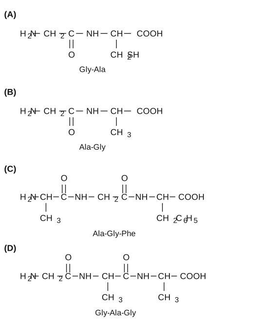
- COMEDK 2024Set 2024-A1 markMCQQ.Among the following peptides, identify the pair where the name and its structure are correctly matched. (A) (B) (C) (D)
›Reveal solutionSolution
The key is to match the side‑chain sequence in the condensed structure with the standard three‑letter amino‑acid abbreviations (N‑terminal to C‑terminal). Only option (C) correctly names the tripeptide Ala‑Gly‑Phe.
Concept & Intuition
Peptide names are written from the N‑terminal (free amino end) to the C‑terminal (free carboxyl end). Each three‑letter code corresponds to a specific side chain:
- Gly (glycine): side chain = H (i.e., the α‑carbon is –CH₂–).
- Ala (alanine): side chain = –CH₃.
- Phe (phenylalanine): side chain = –CH₂C₆H₅ (benzyl).
To check correctness, read the structure from left (N‑terminus) to right (C‑terminus) and list the side chains in order. Then compare that sequence to the name.
Step‑by‑step reasoning
-
Option (A):
Structure: H₂N–CH₂–C(=O)–NH–CH(–CH₂SH)–COOH
- First residue: α‑carbon has no side chain (just –CH₂–) → Gly.
- Second residue: α‑carbon has –CH₂SH (cysteine side chain) → Cys, not Ala. Name given: “Gly‑Ala” → mismatch (should be Gly‑Cys). ✗ Incorrect.
-
Option (B):
Structure: H₂N–CH₂–C(=O)–NH–CH(–CH₃)–COOH
- First residue: –CH₂– → Gly.
- Second residue: –CH₃ → Ala. Name given: “Ala‑Gly” → order reversed (N‑terminal is Gly, not Ala). ✗ Incorrect.
-
Option (C):
Structure: H₂N–CH(–CH₃)–C(=O)–NH–CH₂–C(=O)–NH–CH(–CH₂C₆H₅)–COOH
- First residue: –CH₃ → Ala.
- Second residue: –CH₂– (no side chain) → Gly.
- Third residue: –CH₂C₆H₅ → Phe. Name given: “Ala‑Gly‑Phe” → matches exactly. ✓ Correct.
-
Option (D): …
- COMEDK 2024Set 2024-M1 markMCQQ.Given below are 4 statements about Insulin. Which of these statement/(s) is/are correct? [A] Insulin is a globular protein consisting of 51 Amino acids. [B] Insulin is constituted of 3 polypeptide chains linked together. [C] Insulin is constituted of 2 polypeptide chains linked together by disulphide bonds. [D] The 3 polypeptide chains in Insulin are linked together by Hydrogen bonding. (A) [D] (B) [A] & [C] (C) [B] & [D] (D) [B]
›Reveal solutionSolution
Insulin is a globular protein with 51 amino acids arranged in two polypeptide chains (A and B) linked by disulphide bonds, so only statements [A] and [C] are correct.
Concept and Intuition
Insulin is a classic example of a small, well-studied protein. Its structure is fundamental in biochemistry: it is synthesized as a single chain (proinsulin) that is later cleaved to remove a connecting peptide (C-peptide), leaving two chains — the A chain (21 amino acids) and the B chain (30 amino acids). These two chains are held together by disulphide bonds (covalent bonds between cysteine residues), not by hydrogen bonds between separate chains. Hydrogen bonds are important for the protein’s three-dimensional folding, but they do not link the two chains together. Knowing this, we can evaluate each statement.
Step-by-step reasoning
-
Statement [A]: “Insulin is a globular protein consisting of 51 Amino acids.”
- Insulin is indeed a globular protein (soluble, roughly spherical shape). The total number of amino acids in the active form is 21 (A chain) + 30 (B chain) = 51.
- This statement is correct.
-
Statement [B]: “Insulin is constituted of 3 polypeptide chains linked together.”
- Mature insulin has only two chains (A and B). The third chain (C-peptide) is present only in the precursor proinsulin and is removed during processing.
- This statement is incorrect.
-
Statement [C]: “Insulin is constituted of 2 polypeptide chains linked together by disulphide bonds.”
- Exactly right: the A and B chains are connected by two interchain disulphide bonds (between A7–B7 and A20–B19). There is also one intrachain disulphide bond within the A chain (A6–A11).
- This statement is correct.
-
Statement [D]: “The 3 polypeptide chains in Insulin are linked together by Hydrogen bonding.” …
-
- KCET 2023Set B-41 markMCQQ.Match List-I and List-II with respect to proteins and their functions and select the correct option. List-I | List-II
- Collagen | p. Fights infectious agents
- Trypsin | q. Hormone
- Insulin | r. Enzyme
- Antibody | s. Intercellular ground substance (A) 1-s, 2-p, 3-r, 4-p (B) 1-q, 2-r, 3-q, 4-s (C) 1-s, 2-q, 3-r, 4-p (D) 1-s, 2-r, 3-q, 4-p
›Reveal solutionSolution
Assign each protein its biological role: collagen → ground substance, trypsin → enzyme, insulin → hormone, antibody → defence.
Concept — Proteins and their functions (Biomolecules). NCERT gives a table of proteins with their functions; each protein here has exactly one role.
-
Collagen — the most abundant protein in the animal world; it is the fibrous protein of the intercellular ground substance of connective tissue. → s
-
Trypsin — a proteolytic enzyme secreted (as trypsinogen) by the pancreas; it hydrolyses proteins in the small intestine. → r
-
Insulin — a peptide hormone secreted by the β-cells of the islets of Langerhans; it lowers blood glucose. → q
-
Antibody — an immunoglobulin that fights infectious agents (antigens/pathogens). → p …
- KCET 2019Set A-11 markMCQQ.Which of the following is generally water insoluble? (A) Fibrous protein (B) Amylose (C) Vitamin-C (D) Glycine
›Reveal solutionSolution
The key is solubility in water: fibrous proteins are structural and hydrophobic, while amylose, vitamin-C, and glycine are all water-soluble. The insoluble one is fibrous protein.
Concept & Intuition
Solubility in water depends on how well a molecule can interact with water molecules through hydrogen bonding or ionic interactions. Water is polar, so "like dissolves like" — polar and charged substances tend to dissolve, while nonpolar, hydrophobic ones do not.
Let's look at each option:
-
Fibrous protein — These are structural proteins (like collagen, keratin, elastin) that form long, sheet-like or rope-like fibers. Their amino acid sequences are rich in hydrophobic residues, and they are tightly packed with extensive cross-linking. They are designed to be insoluble in water, providing mechanical strength to tissues. They do not form colloidal solutions in water; they remain as solid fibers.
-
Amylose — This is a linear polymer of glucose (a polysaccharide), a component of starch. Each glucose unit has many –OH groups, which form hydrogen bonds with water. Amylose is actually water-soluble (it forms a colloidal solution in hot water, and even in cold water it disperses to some extent). So it is not generally insoluble.
-
Vitamin-C (ascorbic acid) — This is a small, polar molecule with multiple –OH groups and a lactone ring. It is highly water-soluble. In fact, it is one of the most water-soluble vitamins. Definitely not insoluble.
-
Glycine — The simplest amino acid. It exists as a zwitterion in water (X+X22+HX3N−CHX2−COOX−), making it highly polar and very soluble in water. It dissolves readily.
Watch outA common mistake is to think that all proteins are water-soluble. Many globular proteins (like enzymes, hemoglobin) are soluble, but fibrous proteins are specifically designed to be insoluble — that's their biological function. …
-
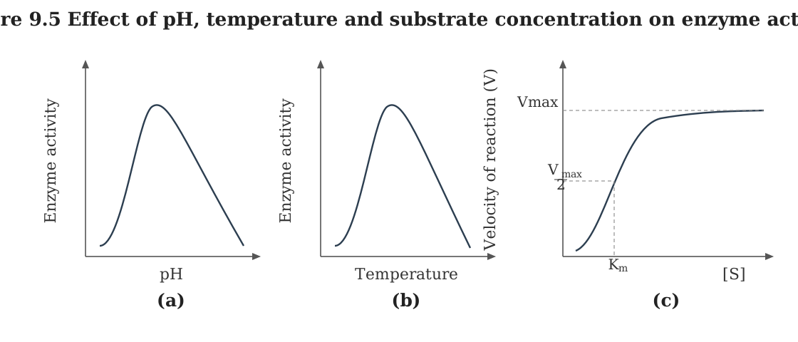
- KCET 2018Set A-11 markMCQQ.The following graph shows concentration of substrate on enzyme activity: (see the graph described below) What does the Y-axis represent?
 (A) Temperature
(B) Velocity of reaction
(C) pH
(D) Pressure
(A) Temperature
(B) Velocity of reaction
(C) pH
(D) Pressure›Reveal solutionSolution
The plateau is labelled Vmax, and V stands for velocity — so the Y-axis is the velocity (rate) of the reaction.
Step 1 — Recognise the curve. A plot of substrate concentration against enzyme activity that saturates at a maximum is the Michaelis–Menten hyperbola:
v=Km+[S]Vmax[S].
Step 2 — Read the axes.
- X-axis = substrate concentration [S] (stated in the stem).
- Y-axis = the velocity (rate) of the reaction v; it rises steeply at low [S] and asymptotes to the horizontal line marked Vmax, the maximum velocity reached when every active site is occupied. …
- KCET 2018Set A-11 markMCQQ.The primary gases that were used by Miller in his experiment are (A) CH4,NH3,H2O,H2 (B) CH4,CO2,N2,SO2 (C) CH4,CO2,N2,NH3 (D) CH4,N2,NH3,H2
›Reveal solutionSolution
Miller's spark-discharge experiment used a reducing atmosphere: CH4, NH3, H2 and water vapour.
Step 1 — The hypothesis being tested.
Oparin and Haldane proposed that the first organic molecules formed abiotically from simple inorganic molecules in a reducing (oxygen-free, hydrogen-rich) primitive atmosphere, with energy supplied by lightning and heat. Miller set out to test exactly this.
Step 2 — The apparatus and the gases.
In 1953 S. L. Miller built a closed flask containing:
- CH4 (methane) — the carbon source
- NH3 (ammonia) — the nitrogen source
- H2 (hydrogen) — makes the mixture strongly reducing
- water vapour (H2O) — from a flask of boiling water
Electric discharge (~800 °C, simulating lightning) was passed through the mixture.
Step 3 — Why the other options fail. …
🎓Unlock everything free for 14 days
- ✓Full step-by-step solutions
- ✓Concept-first explanations
- ✓Methods, shortcuts & mistakes
- ✓PYQ mapping + timed mock tests
Full access for 14 days. No credit card required.