Q.Observe the diagram. Answer the question.
You're viewing a preview — the full solution, concept, methods & PYQ mapping are locked.
Start your 14-day free trial to unlock the full solution →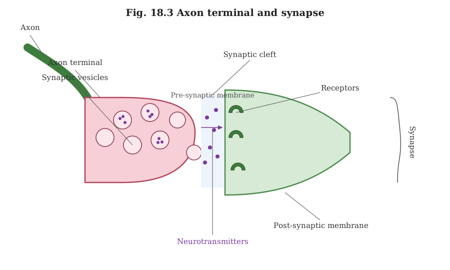
The figure is a chemical synapse: the axon terminal bulb (with its cleft/gap, vesicles, and neurotransmitter release site) meeting the postsynaptic membrane and its receptors.
A chemical synapse consists of a knob-like swelling at the end of the presynaptic axon terminal, packed with membrane-bound sacs called synaptic vesicles containing neurotransmitter molecules, separated from the next neuron's membrane by a narrow fluid-filled gap (the synaptic cleft).
Based on the figure's described layout — the wedge-shaped presynaptic terminal bulging into vesicle-filled swelling, with an outer membrane/gap region at upper right, a large vesicle, a row of vesicles near the lower membrane, and a lower membrane bearing small tick-mark (receptor) symbols, with "Neurotransmitters" labelled near the membrane at the bottom:
- (a) — pointing to the outer membrane/cleft region — is the synaptic cleft, the narrow gap between the presynaptic and postsynaptic membranes across which neurotransmitter diffuses.
- (b) — pointing to one of the larger circular vesicles — is a synaptic vesicle, a membrane-bound sac storing neurotransmitter molecules within the axon terminal.
- (c) — pointing to the row of vesicles clustered along the lower (presynaptic) membrane — represents synaptic vesicles docked at the presynaptic membrane, ready to fuse with it and release their neurotransmitter into the cleft by exocytosis. …
Unlock everything free for 14 days
- Full step-by-step solutions
- Concept-first explanations
- Methods, shortcuts & mistakes
- PYQ mapping + timed mock tests
Full access for 14 days. No credit card required.