Q.Define mitosis. Describe the different phases of mitosis with well labelled diagrams. OR With a labelled diagram describe the internal structures of a dorsiventral leaf.
You're viewing a preview — the full solution, concept, methods & PYQ mapping are locked.
Start your 14-day free trial to unlock the full solution →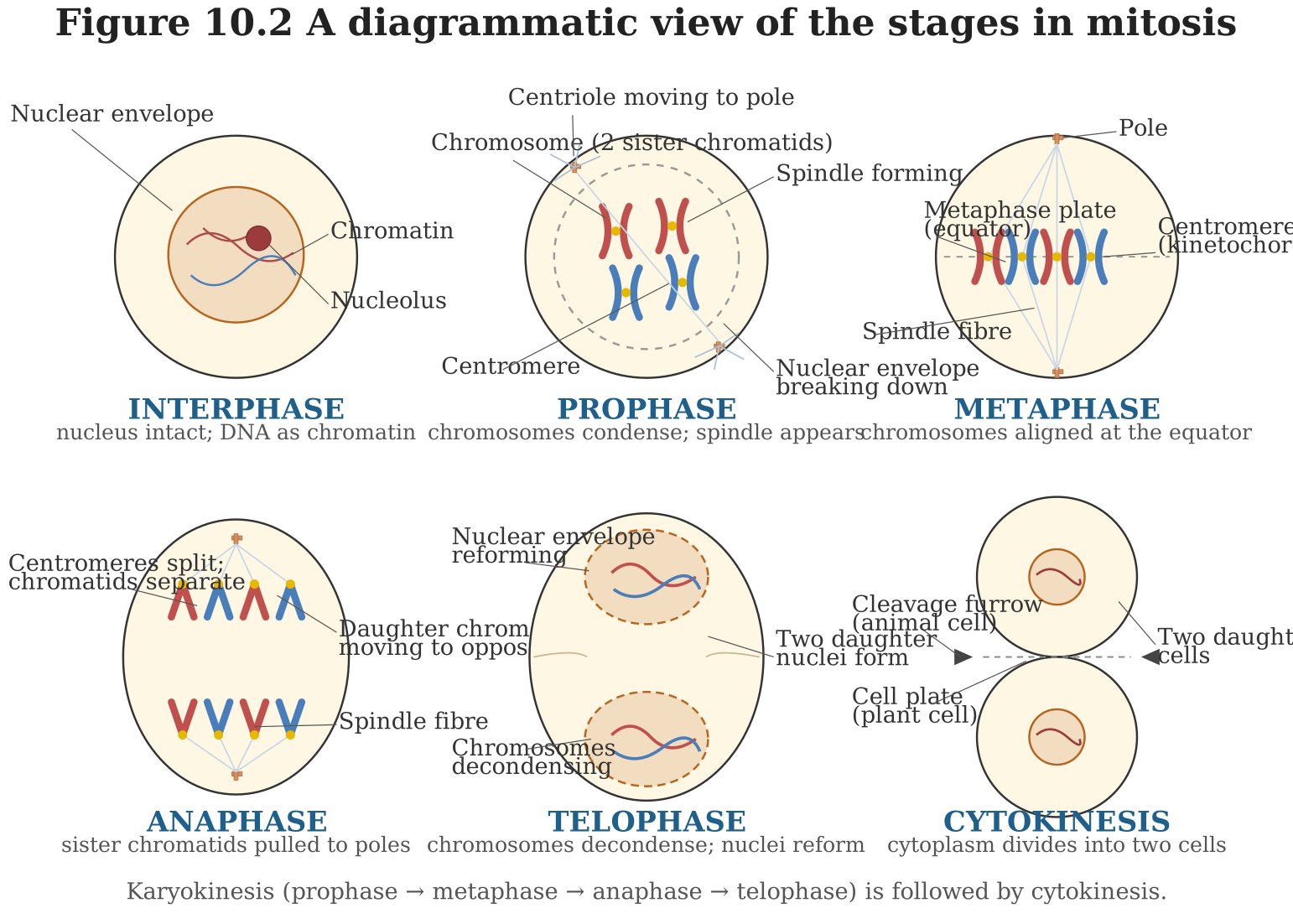
Mitosis is equational (somatic) cell division producing two genetically identical daughter cells, occurring through four main phases — prophase, metaphase, anaphase and telophase — followed by cytokinesis.
This is a core topic of the Cell Cycle and Cell Division chapter. Since a rendered diagram cannot be produced here, each phase is described in full written detail, in the order it would be shown and labelled on a real diagram.
Definition: Mitosis is a type of cell division (also called equational division) in which a single (diploid) parent cell divides once to produce two daughter cells, each genetically identical to the parent cell and to each other, with the same chromosome number as the parent. It is the mode of division responsible for growth, repair and replacement of cells in the body (somatic cell division).
The M Phase (Mitosis) is divided into four sub-phases:
1. Prophase: (Diagram description: a cell with chromatin condensing into visible, thread-like chromosomes, each consisting of two sister chromatids joined at the centromere; the nucleolus and nuclear envelope beginning to disappear; centrioles (in animal cells) moving to opposite poles with spindle fibres starting to form between them.) — The chromosomal material (chromatin) condenses to form compact, visible chromosomes. Each chromosome is seen to consist of two sister chromatids held together by a centromere. The nucleolus gradually disappears, and the nuclear envelope begins to break down. Centrioles move towards opposite poles of the cell, and spindle fibres begin to form.
2. Metaphase: (Diagram description: the nuclear envelope fully disappeared; all chromosomes arranged in a single row at the exact centre/equator of the cell, each attached to spindle fibres from both poles via its centromere/kinetochore.) — The nuclear envelope is now completely disintegrated. The chromosomes, each still with its two sister chromatids, align themselves at the equatorial plate (metaphase plate) of the cell, attached to spindle fibres from opposite poles via their kinetochores. This precise alignment ensures that each daughter cell will receive an identical set of chromosomes.
3. Anaphase: (Diagram description: the centromere of each chromosome splitting, with the two sister chromatids (now individual chromosomes) being pulled apart and moving towards opposite poles of the cell along the spindle fibres, giving a characteristic V or J shape.) — The centromere joining each pair of sister chromatids splits, and the sister chromatids separate, becoming independent chromosomes. These are then pulled towards opposite poles of the cell by the shortening spindle fibres.
4. Telophase: (Diagram description: chromosomes having reached the two poles, decondensing back into diffuse chromatin; a new nuclear envelope forming around each set at either pole; nucleolus reappearing; a cell plate/cleavage furrow beginning to form at the centre for cytokinesis.) — At each pole, the chromosomes decondense and revert to their extended chromatin form. A new nuclear envelope reforms around each group of chromosomes, forming two daughter nuclei, and the nucleolus reappears in each. This is usually followed immediately by cytokinesis — division of the cytoplasm — via a cleavage furrow (in animal cells) or a cell plate (in plant cells), resulting in two separate, genetically identical daughter cells.
…
Unlock everything free for 14 days
- Full step-by-step solutions
- Concept-first explanations
- Methods, shortcuts & mistakes
- PYQ mapping + timed mock tests
Full access for 14 days. No credit card required.