Question of 48
Q.Draw a labelled diagram of a myofibril of skeletal muscle showing different region.
Meghalaya MboseMBOSE Meghalaya 11th Board 2019Subjective· 3mImportance★★★★★
0% · 0/48 Questions
You're viewing a preview — the full solution, concept, methods & PYQ mapping are locked.
Start your 14-day free trial to unlock the full solution →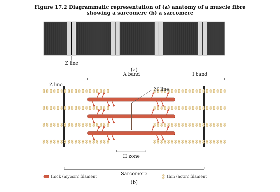
Each myofibril is made of repeating sarcomeres — the region between two Z lines — showing alternating I bands (thin actin filaments) and A bands (thick myosin filaments overlapping actin), with an H zone and M line at the centre.
Since a hand-drawn image cannot be produced here, the structure of one sarcomere (the repeating unit of a myofibril) is described region by region, as it would be labelled on a diagram:
- Z line: a dense line marking the boundary of each sarcomere; thin (actin) filaments are anchored here. The sarcomere is defined as the region between two successive Z lines.
- I band (light/isotropic band): the region containing only thin actin filaments, appearing lighter under the microscope; it is bisected by the Z line.
- A band (dark/anisotropic band): the region spanning the full length of the thick myosin filaments, appearing darker; in the resting sarcomere it includes the zone where actin and myosin filaments overlap.
- H zone: a lighter zone at the centre of the A band, where only thick myosin filaments are present (no overlapping actin), visible in a relaxed muscle. …
Unlock everything free for 14 days
- Full step-by-step solutions
- Concept-first explanations
- Methods, shortcuts & mistakes
- PYQ mapping + timed mock tests
Full access for 14 days. No credit card required.