Physical Education · Ch 7 — Physiology and Injuries in Sports
Hard Tissue Injuries
Hard Tissue Injuries
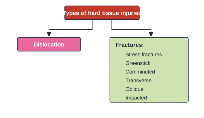
An injury to the skeletal system of the body — one in which a bone cracks or breaks — is called a hard tissue injury. Hard tissue injuries are of two kinds: dislocation and fractures.
Drawn by us to help you understand the concept clearly, and verified to make sure it's accurate. For exams, practice from your textbook's own diagram.
A tree/flow diagram. A parent box labelled 'Types of hard tissue injuries' branches to two boxes: 'Dislocation' on one side, and 'Fractures:' on the other listing six fracture types — Stress fractures, Greenstick, Comminuted, Transverse, Oblique and Impacted. (Draw the correct spelling 'Comminuted'; …
Dislocation
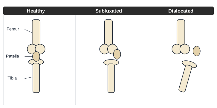
A dislocation is a joint injury that forces the ends of bones out of position. Also called a luxation, it occurs when there is an abnormal separation at a joint, where two or more bones meet; a partial dislocation is called a subluxation. The cause is often a fall or a blow — sometimes from a contact sport — or the weakening of the muscles and tendons around the joint. A dislocated joint can be treated through medication, manipulation, rest or surgery.
Drawn by us to help you understand the concept clearly, and verified to make sure it's accurate. For exams, practice from your textbook's own diagram.
An own-drawn, simplified three-panel comparison of a knee joint (not the textbook's third-party image). Panel 1 'Healthy' — femur, patella and tibia in normal alignment; Panel 2 'Subluxated' — the bones partially displaced (partial dislocation); Panel 3 'Dislocated' — the bone ends fully forced out of position. La …
- Causes — Trauma that forces a joint out of place. Accidents, falls and contact sports such as football are common causes. Dislocations can also occur during ordinary activities when the muscles and tendons around a joint are weak, which is why they happen more often in older people with weaker muscles and balance problems.
- Symptoms — These vary with the severity and location of the injury, and include pain, swelling, bruising, instability of the joint, loss of the ability to move the joint, and a visibly deformed joint (the bone looks out of place).
- Treatment — This depends on the severity and the joint involved. Applying ice and keeping the joint elevated helps reduce pain while waiting to see a doctor. Options include medication for pain, manipulation (a doctor returns the bones to their proper places), rest (protecting and immobilising the joint with a sling or splint so it can heal), rehabilitation (physical-therapy exercises that strengthen the surrounding muscles and ligaments), and surgery — needed if manipulation fails to reset the bones, or if the dislocation has damaged blood vessels, nerves, bones, muscles or ligaments that require repair.
Fractures
A fracture is a break in a bone. Most fractures are caused by a direct impact, such as a fall or a severe tackle, while stress fractures build up over time from overuse. The syllabus covers the following types.
Stress fracture
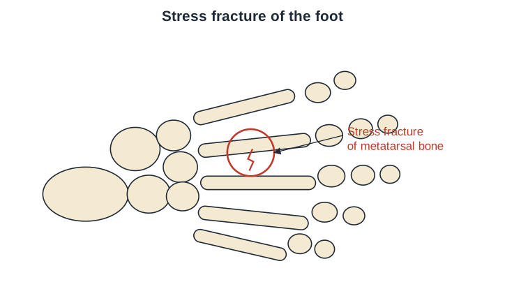
Stress fractures can occur because of overuse injuries and a lack of adequate equipment to protect the body.
Drawn by us to help you understand the concept clearly, and verified to make sure it's accurate. For exams, practice from your textbook's own diagram.
An own-drawn line diagram of the bones of a foot (tarsals, metatarsals and phalanges). A hairline crack is marked on one metatarsal bone and circled, labelled 'Stress fracture of metatarsal bone'. Unlike the dramatic break shown in a transverse or oblique fracture, a stress fracture is a fine, thread-like crack caused by repeated loading rather than a single impact — a reminder that overuse injuries from repetitive running or jumping can be just as serious …
- Causes — Often the result of increasing the amount or intensity of an activity too quickly.
- Prevention — Add low-impact activities to the exercise routine to avoid repeatedly stressing one part of the body.
- Treatment — Rest, cold therapy (ice packs and cold compresses applied to the injured area) and anti-inflammatory medication such as ibuprofen or aspirin; a recovery time of about 6 to 8 weeks is usually needed for healing.
Greenstick
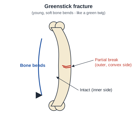
A greenstick fracture is a fracture in a young, soft bone in which the bone bends rather than breaking all the way through.
Drawn by us to help you understand the concept clearly, and verified to make sure it's accurate. For exams, practice from your textbook's own diagram.
An own-drawn schematic (not the textbook X-ray). A long bone (such as a child's forearm bone) shown bent/bowed, with a partial crack on the outer, convex side while the inner side stays intact — like a bending green twig. Labels: 'Bone bends', 'Partia …
- Causes — These fractures most commonly occur with a fall.
- Prevention — Encourage regular exercise, ensure the child's safety with proper protective equipment, and provide adequate calcium in the child's diet.
- Treatment — Removable splints give better outcomes than casting in children (for example, torus fractures of the distal radius).
Comminuted
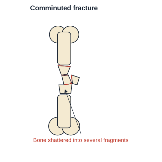
A comminuted fracture is one in which a bone is broken, splintered or crushed into a number of pieces.
Drawn by us to help you understand the concept clearly, and verified to make sure it's accurate. For exams, practice from your textbook's own diagram.
An own-drawn schematic (not the textbook X-rays). An upright long bone broken and crushed into three or more separate fragments at the mid-shaft, labelled 'Bone shattered into several fragments'. A student should read this as the most severe fracture pattern in the chapter: instead of one clean break line, the bone has split into several loose pieces, which is exactly why treatment needs an open reduction — a surgeon must physically rejoin multiple fr …
- Causes — Direct or indirect trauma or violence.
- Prevention — Keep bones strong by eating calcium-rich food and exercising regularly.
- Treatment — An X-ray is important for diagnosis. An open reduction, in which the bone fragments are joined together with surgical nails, wires or plates, is usually required. (The textbook's own diagram misspells this type as "Commutated"; the correct term is comminuted.)
Transverse
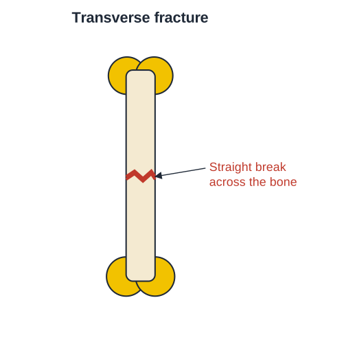
A transverse fracture is a straight break running right across a bone.
Drawn by us to help you understand the concept clearly, and verified to make sure it's accurate. For exams, practice from your textbook's own diagram.
An own-drawn schematic of a single upright long bone with rounded joint ends and one straight, horizontal break running right across the mid-shaft, labelled 'Straight break across the bone'. The break line sits at a right angle to the long axis of the bone — the single feature that identifies a transverse fracture and distinguishes it at a glance from the diagonal break line of an oblique fracture shown in the next figure. This straight p …
- Causes — When a large force is transmitted directly — that is, perpendicularly — to the bone.
- Prevention — Weight-bearing physical activity makes bones stronger and denser; a calcium-rich diet and regular exercise help too.
- Treatment — Can often be managed at home with rest and medicine. A back brace (called a TSL) or an abdominal binder may be prescribed to reduce pain by limiting motion at the fracture site.
Oblique
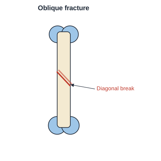
An oblique fracture is one in which the bone breaks diagonally.
Drawn by us to help you understand the concept clearly, and verified to make sure it's accurate. For exams, practice from your textbook's own diagram.
An own-drawn schematic of a single upright long bone with rounded joint ends and one diagonal (slanting) break across the shaft, labelled 'Diagonal break'. The slanted break line is the key visual difference from the transverse fracture in the previous figure: where that break runs straight across at a right angle to the bone, this one cuts across at an angle, typically the re …
- Causes — Usually an injury to the bone from a fall, accident or other trauma.
- Prevention — Strengthen bones by eating calcium-rich food and exercising regularly. …
Drawn by us to help you understand the concept clearly, and verified to make sure it's accurate. For exams, practice from your textbook's own diagram.
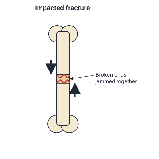
An own-drawn schematic of a single upright long bone broken at the mid-shaft, where the two broken ends are driven and jammed into one another. Two arrows point toward each other at the break, labe …